POLYMER COATINGS TO CONTROL IN-STENT RESTENOSIS
Biomer and Liverpool University
Stent surgery is routine these days: the insertion of a metal framework inside a narrowed blood vessel ensures that the blood can flow properly. However, there are drawbacks: stents can encourage the growth of smooth muscle cells, which can lead to further blockages. Researchers from Biomer Technology, which develops polymers for medical devices, have collaborated with Rachel Wilson, a biocompatibility expert from Liverpool University, to develop a polymer that would prevent this from happening.
The subject of a Knowledge Transfer Partnership, the project brought together Biomer’s materials and chemistry with Wilson’s cell-culture testing to establish an in-vitro test to determine whether stents — as well as other cardiovascular devices — would be biocompatible.
The key property for the stents is that they inhibit the growth of smooth muscle cells and stimulate the growth of endothelial cells, which form the lining of healthy blood vessels. The project, according to Biomer, has allowed it to identify candidate materials for biocompatible polymer applications earlier and to speed up their optimisation. This has allowed it to get its products into clinical trials earlier; one new polymer is currently in ‘firstin- human’ trials in the US.

DEVELOPMENT OF LARGE BEARING HIP REPLACEMENTS
Finsbury Development and Southampton University
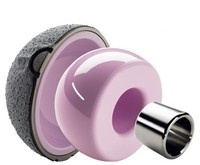
Bone implants, such as hip joints, have to withstand extremely tough conditions — constant large loading forces in a corrosive environment. The bioengineering research group at Southampton University’s school of engineering sciences has developed a hip joint that uses tough ceramics to give a wider range of motion than has previously been possible, while reducing the risk of dislocation.
The concept for the new joint was a thin ceramic insert within a titanium cup that would integrate with the surrounding bone.
Although ceramics are highly resistant to the conditions inside the body, the insert required for these implants has to be extremely thin. As ceramics also tend to be brittle, it has not been possible up until now to make them thin enough, or accurately enough, to work well as hip-joint bearings.
Working with Finsbury Orthopaedics as part of a Knowledge Transfer Partnership, the Southampton team used design techniques from the aerospace sector — probabilistic ceramic design and analysis — to optimise the thickness of the ceramic shell. It also developed a pre-assembly technique for putting the implant together that reduces the risk of it coming apart after implantation, as well as a model that predicts the response of the surrounding bone to the implant. The result is an implant that uses a ceramic head within a surrounding socket in an anatomically correct configuration, which should wear more slowly than previous implants and which requires less follow-up surgery. So far, 200 implants have been carried out around the world.
CYMAP
Gray Institute for Radiation Oncology and Biology, The Technology Partnership, Oxford University and Cancer Research Technology
Medical imaging has found ways to see many of the structures within the human body, but one that has always caused problems is the cell. Taking off from a UK Research Councils project to develop ‘optical biochips’ to identify, locate and monitor changes in cells, the Gray Institute for Radiation Oncology and Biology (GIROB) has developed a system that departs radically from the usual approach to imaging.
Rather than relying on reflected light to provide an image, GIROB’s technique, which is called CyMap, works by exploiting one of the properties of cells that makes them hard to see: they are transparent. When light passes through cells, however, it is diffracted and imaging electronics, such as charge-coupled devices, can detect the diffraction patterns.
If the cell’s state changes, the shape of the diffraction pattern also changes. As the system uses diffraction rather than reflection, it does not require a lens to focus on the subject; in fact, it does not use a lens at all.
Among the most exciting applications for CyMap are its use as a low-cost, portable diagnostic tool; it could detect the presence of pathogenic bacteria in blood samples in the developing world and could also be used to test drinkingwater purity.


April 1886: the Brunkebergs tunnel
First ever example of a ground source heat pump?