
It uses electromagnets instead of radiation-based imaging, researchers report in the American Heart Association journal Circulation: Arrhythmia and Electrophysiology.
Electromagnetic tracking technology was used successfully to implant Cardiac Resynchronisation Therapy (CRT) devices in heart failure patients, and cut patients’ and doctors’ radiation exposure as compared with traditional fluoroscopy-based implantation procedures.
CRT is becoming more common as a treatment option for patients with severe congestive heart failure and conduction disease. Beyond conventional right ventricular pacing, a CRT device, which provides biventricular pacing, helps the left and right lower chambers of the heart to beat in synchrony. Although fluoroscopy is traditionally used to guide CRT implantation, radiation exposure from this type of imaging is a major concern even for experienced operators using an up-to-date scanner.
The new tracking system uses tools equipped with sensors that can be detected electromagnetically, which reduces the need for conventional fluoroscopy.
Average fluoroscopy time for traditional CRT implantation exceeds 20 minutes but with the new tracking system, patients were exposed to the conventional fluoroscopy an average of five minutes.
‘This new technology has the potential to revolutionize the way we image inside the body while we perform a wide range of diagnostic and therapeutic interventions in the future,’ said the study’s lead author Sergio Richter, M.D., consulting electrophysiologist and senior physician at the Heart Center of the University of Leipzig in Germany.
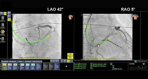
Researchers have compared the new electromagnetic navigation system to a GPS tracking system. ‘Just as data from three satellites orbiting the earth are used to locate a GPS device’s location on earth, the MediGuide system uses data from three transmitters to measure a sensor’s position and orientation within the electro-magnetic field around the patient at all times,’ Richter said in a statement.
Unlike conventional 2D fluoroscopy, this technology applies three-dimensional visualisation to pre-recorded 2D images used by the physician for CRT implantations. This creates a real-time clinical environment that adjusts automatically for heart rate, respiratory motion and patient motion, and accurately tracks catheter position within 1mm and 1 degree, Richter said.
The new tracking system was used in 15 heart failure patients (eight men and seven women, average age 66) who entered Leipzig University Hospital early in 2012 and met the American College of Cardiology/American Heart Association/Heart Rhythm Society indications for CRT implantation. Study patients were initially followed for four weeks after implantation. All 15 patients were identified as severe heart failure patients.
Traditional fluoroscopy-guided implantation was used for the two device leads on the right side of the heart. The new tracking technology was used to place the third device lead via the coronary sinus on the left ventricle, the heart’s largest and main pumping chamber.
‘Patients with higher body mass and those requiring a large number of fluoroscopy-based interventions, either diagnostic or therapeutic, such as chronic disease or cancer patients, would especially benefit as they are at a higher risk of increased cumulative exposure to harmful radiation and to higher risk of developing new or secondary cancers,’ Richter said.


Swiss geoengineering start-up targets methane removal
Several rather dubious statistics in this report. IF methane had 120× the thermal effect of CO2 that would be TWO orders of magnitude. Two is not...