
The titanium dioxide nanoparticles are applied to the material or object to be viewed, rather than being fixed to the microscope. Droplet-like lens structures formed of millions of nanobeads break up light, refracting it to illuminate objects with tiny individual beams. According to the study, published in the journal Science Advances, the superlens adds 5x magnification to existing microscopes.
“We've used high-index titanium dioxide (TiO2) nanoparticles as the building element of the lens,” said Dr Zengbo Wang from Bangor University. “These nanoparticles are able to bend light to a higher degree than water. To explain, when putting a spoon into a cup of this material, if it were possible, you'd see a larger bend where your spoon enters the material than you would looking at the same spoon in a glass of water."
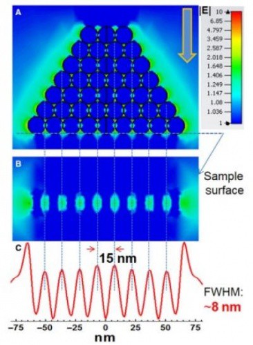
"Each sphere bends the light to a high magnitude and splits the light beam, creating millions of individual beams of light. It is these tiny light beams which enable us to view previously unseen detail."
The physical laws of light mean that objects smaller than 200nm can’t be viewed with a normal microscope. Using the superlens, however, the team was able to view certain details on objects for the first time, including the minuscule data grooves on the surface of a Blue Ray DVD. In addition, the researchers say that titanium dioxide is cheap and readily available, and that other labs around the world should be able to replicate the technology and adapt it for their own use.
"We have already viewed details to a far greater level than was previously possible,” said Wang. “The next challenge is to adapt the technology for use in biology and medicine. This would not require the current use of a combination of dyes and stains and laser light- which change the samples being viewed. The new lens will be used to see germs and viruses not previously visible."


Red Bull makes hydrogen fuel cell play with AVL
Formula 1 is an anachronistic anomaly where its only cutting edge is in engine development. The rules prohibit any real innovation and there would be...