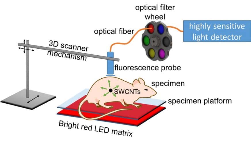
The method, described in the Royal Society of Chemistry journal Nanoscale, relies on spectral triangulation. Cancerous tumours are tagged with antibody-linked nanotubes, which naturally fluoresce at short-wave infrared wavelengths when excited by visible light. A highly sensitive detector called an InGaAs (indium gallium arsenide) avalanche photodiode allows for the faint signals from the nanotubes to be identified up to 20mm deep in the simulated tissue used for testing.
"We're using an unusually sensitive detector that hasn't been applied to this sort of work before," said Rice chemist Bruce Weisman, who led the research. "This avalanche photodiode can count photons in the short-wave infrared, which is a challenging spectral range for light sensors. The main goal is to see how well we can detect and localise emission from very small concentrations of nanotubes inside biological tissues. This has potential applications in medical diagnosis."
The Rice team used LED lighting to excite the nanotubes. According to Weisman, lasers are more commonly used for this, but can’t be focused inside tissue because of scattering. However, LED light diffuses through tissue and is able to penetrate to the nanotubes.
A small optical probe mounted on the frame of a 3D printer follows a programmed pattern, as the probe gently touches the skin to make readings at grid points spaced a few millimetres apart. Water in the tissue absorbs different wavelengths of nanotube emission, meaning the team is able to establish the depth of a signal, as well as the X and Y coordinates.
"If we're detecting nanotubes close to the surface, the long and the short wavelength emissions are relatively similar in intensity,” said Weisman. "But if the emission source is deeper, water in that tissue absorbs the longer wavelengths preferentially to the shorter wavelengths. So the balance between the intensities of the short and long wavelengths is a yardstick to measure how deep the source is. That's how we get the Z coordinate."
The detector is currently being tested by Dr Robert Bast, an ovarian cancer expert and research academic at the University of Texas MD Anderson Cancer Centre.


April 1886: the Brunkebergs tunnel
First ever example of a ground source heat pump?