We’re accustomed to engineering skills playing a central role in answering some of the big physics questions about the universe and the subatomic world. But new research using methods honed in plasma fusion reactors could address one of the central problems in biology: namely how the human brain functions at a mechanistic level.
Of course, we already have useful scanning techniques such as functional magnetic resonance imaging (fMRI) that give nice images showing different areas of the brain lighting up when subjects are asked to perform certain tasks. But fMRI is not without some major limitations: it is an indirect measure looking at blood oxygen perfusion as a proxy for actively firing neurons; and it frequently requires a bit of creative statistics to make the science stick.
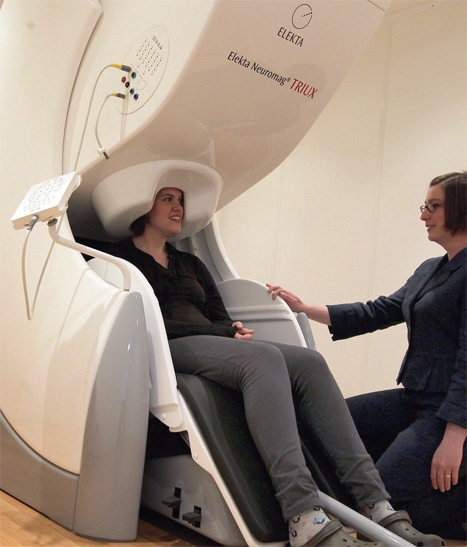
Magnetoencephalography (MEG) is a scanning technique that is, in many ways, the opposite of MRI. Rather than using huge coils to generate magnetic fields of several Tesla and seeing how the brain reacts, MEG passively observes the tiny, femto-Tesla fields naturally generated by firing neurons. The technique has been around for more than 10 years and has found a practical use in surgical planning for removing brain lesions (see panel).
More recently, though, MEG has attracted attention as a research tool, coinciding with a school of thought in neuroscience that tries to understand the brain using a network approach.
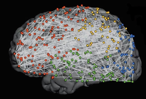
One such advocate is Prof Ed Bullmore, a neuroscientist at Cambridge University. ‘Our brains have billions of nerve cells connected by trillions of synapses, so trying to figure out how it works by focusing on one cell or one synapse at a time is impossible,’ he said at last year’s Cambridge Science Festival. ‘But we can use some simple mathematics to give us a different vision of the brain - losing sight of many of the details but clarifying the complex overall pattern of connections that make up a brain network.’
Saying that the brain is complex might seem a bit obvious, but ‘complex systems’ is actually a radical new cross-disciplinary branch of science that seeks to understand how parts of a system give rise to the collective behaviours of the whole and how the system interacts with its environment.
A simple model is that of an avalanching sand pile. If you drop grains of sand on top of a pile, it will grow in a way that is fairly easy to model. Occasionally, a new grain of sand will trigger an avalanche, but it is very difficult to predict if the next grain of sand will cause an avalanche, where that avalanche will occur and how many grains of sand will be involved.
A key feature of complex systems is that they are neither completely random nor completely ordered.
‘If you record the activity of several different cells simultaneously, it turns out they occasionally show these bursts of synchronised firing - that’s called a neuronal avalanche,’ Bullmore told The Engineer. ‘Those bursts of firing follow a parallel probability distribution, which is the same for an avalanche in the mountains. It’s a metaphor but it does highlight the fact that many statistical properties seem to be conserved from one complex system to another, even if those systems are substantively different from one another.’
“Although not prescriptive, the brain’s response to surprise is considered to be one of the six universal emotional responses, alongside anger, fear, disgust, happiness and sadness”
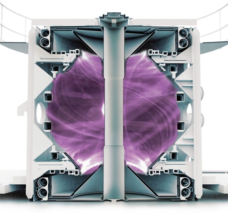
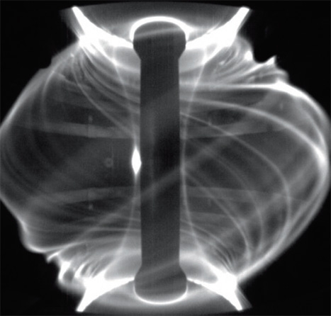
Indeed, the avalanche complex system model is something that caught the attention of another researcher in a very different field. Prof Sandra Chapman of Warwick University studies magnetically confined plasmas for fusion energy, as well as the naturally occurring plasmas in the sun and solar wind (see panel). ‘The problem with fusion is how to keep it hot: it wants to cool, it wants to radiate its heat out, and you’re trying to stop it doing that. If you have turbulence - big fluctuations - it’s bad in a tokomak because the heat can get out more quickly, so it’s trying to understand the physics of the turbulence,’ said Chapman.
Following a conference on complex systems that the two researchers attended, Chapman suggested that some of the data techniques she had developed for maintaining and observing confined plasmas could be useful in interpreting signals from MEG scans, with a view to resolving certain network maps. ‘It’s a very similar problem: you’re making measurements with MEG on the surface of the skull and trying to diagnose what’s going on inside,’ she said. ‘There are currents inside the brain just as in the tokomak there are currents flowing around doing stuff and it’s all very dynamic, but you can’t put the probes into plasma because it’s hot - you have to diagnose it from the edge of the tokomak. Most of my time is spent trying to figure out clever ways of doing this sort of data analysis.’
The problem with MEG is threefold: the signal is of course very small (in fact, a ticking clock generates a magnetic field around a million times stronger); its frequency limit is in the gamma band at around 33-64Hz (not bad by medical scanning standards, but the brain’s network connections can tick over in around 100ms); and finally there is inter-subject variability to consider.
‘You’re looking for the brain’s response to a particular thing, but if you do MEG on a resting person, you don’t get zero - you get all sorts of stuff - because they’re thinking about their cat and each person has a different resting state,’ said Chapman.
The first problem the collaborative team set to work on was characterising the brain’s response to surprise. The research was supported by GlaxoSmithKline, the MRC and by an EPSRC ‘cross disciplinary feasibility account’. Although not prescriptive, surprise is considered one of the six universal emotional responses (alongside anger, fear, disgust, happiness and sadness) and there are robust experimental methods to isolate and study it.
They used something called the mismatch-negativity (MMN) paradigm. While in an MEG scanner, 10 healthy volunteers listened to a series of beeps, some of which were regular and repetitive, and some of which were different and out of sequence.
Previous studies have established that the lateral temporal auditory cortex and frontal cortex are the main brain sites thought to be involved. But none yet have looked at how brain networks respond dynamically. ‘We basically binned the connections into two kinds: one within the different regions, like the temporal lobe, and ones that reach between one and the next. Then we just plotted them and counted them,’ said Chapman. ‘We’re looking at the ratio between lobe connections and the ones within - that’s the new thing.’
After recording the data under this framework, the team had the unenviable task of interpreting the results. However, Chapman’s experience wrestling with plasma came in particularly useful here.
‘If I have a signal at A and a signal at B and I want to see if they are in fact correlated, I have to show that’s not through random processes,’ she said. ‘This is additionally tricky because it’s an integrating measure; the sensors are on the surface and the signal is inside the brain, so you will pick up at more than one sensor if you just look at the amplitude of the signal - the amplitude is useless really - so we look at the phase.’
The team found that when subjects heard the predictable standard tone, the brain tended to synchronise locally with lots of intra-lobe connections. Enter the surprising tone, though, and the number of long-range inter-lobe connections increased while at the same time the number of local connections actually fell, then returned to normal resting state again, all in around 80ms. This was quite surprising, according to Chapman.
‘The expectation was that maybe the brain would just grow more connections when you ask it to do more work,’ she said. ‘You’d always have the local response and you’d add on top of it a whole bunch of extra connections to deal with the surprise, but that’s not quite what’s happening. It has the same number of connections but fewer local ones and more global ones when it has to do something. The brain is trying to be efficient so it’s using fewer local connections and more of these global ones - so it’s dynamically reconfiguring.’
“Many aspects of the brain are organised so as to minimise or nearly minimise wiring cost”
Prof Ed Bullmore, Cambridge
Although in some ways unexpected, this reconfiguring actually fits well with some of Bullmore’s ideas about how brain networks are constrained by certain parameters in the same way any other complex networks are.
To understand this, you first have to appreciate that the brain is ‘metabolically expensive’ - it accounts for only two per cent of the total mass of the human body but 20 per cent of its total energy budget. ‘Many aspects of the brain are organised to minimise or nearly minimise wiring cost,’ said Bullmore. ‘One reason why the brain has such a convoluted, wrinkled appearance is to save wiring cost.’
He believes there is an inherent trade-off in brain networks between wiring cost and the efficiency of information transfer. ‘Normally functioning brains probably renegotiate that trade-off second by second depending on the information processing demands. When you repeatedly present the brain with a rather boring, predictable stimulus, it settles back into a relatively low-cost configuration. It doesn’t spend extra money on long-distance and high-efficiency communication until it’s presented with a surprising stimulus.’
The findings could have particular relevance to complex psychiatric diseases such as schizophrenia, where patients show an inability to filter out unnecessary information. It could also alter the way we think about the brain. ‘[Trade-offs] may be a better way of thinking about how the brain has been selected by evolution, rather than thinking it’s been selected for performance without any cost constraint at all. It’s just like the rest of life really; it’s a question of value for money.’
in depth
getting a reaction
Although cross-disciplinary research usually starts as one field helping out another, the expectation is usually that the favour will be returned at some point.
‘It’s not just that we’ve got these great ideas and we give them to the neuroscientists - they have ways of doing things we can bring back to plasmas that I hadn’t thought of,’ said Prof Sandra Chapman.
Some of these ideas might find their way into the JET and MAST fusion reactors at Culham, where Chapman sometimes works, and eventually ITER - the international fusion facility being built in France.
The common aim for fusion research projects is to help achieve sustained reactions that produce more energy than is put in. Understanding magnetically confined plasmas is a large part of that.
‘Tokamaks, if you persuade them with the right amount of heating or jiggling - this is an empirical thing - they go into an enhanced confinement state called H-mode, but it’s not really understood how exactly,’ explained Chapman. Sustaining this H-mode (which differs from the resting ‘L-mode’) could be the key to net power. However, one problem with H-mode is the occasional appearance of turbulent features called edge-localised modes (ELMs).
‘It would be nice to tell what the plasma is about to do; is it about to go into H-mode or come out of H-mode or is an ELM going to come along? If you get ELMs in ITER, they will knock a hole in the wall, so the idea, between now and then, is to solve this problem.’
in depth
thinking ahead
The MEG technique is currently being used in surgical planning to non-invasively remove brain lesions
It all really starts in the 19th century with James Clerk Maxwell and Michael Faraday demonstrating that any current will generate a magnetic field.
Magnetoencephalography (MEG) is based on arrays of superconducting quantum interference devices (SQUIDs), which exploit the phenomenon of electron-pair wave coherence to achieve the very low noise detection of weak neuromagnetic signals.
Since SQUIDs are based on superconductor technology, they require cryogenic temperatures. In a modern MEG device, an array of more than 300 SQUIDS is contained in a helmet-shaped liquid-helium-cooled vessel called a dewar, allowing simultaneous measurements at many points over the head.
The MEG system is operated in a shielded room that minimises interference from external magnetic disturbances, including the Earth’s magnetic field and objects such as cars and trains.
One of the major current uses of MEG in hospitals is in surgical planning to remove brain lesions. Traditionally, if a patient had a lesion close to the motor cortex or speech and language areas, a surgeon would remove the skull around those areas, wake them up and then stimulate those parts of the brain around the lesion to make sure that he or she doesn’t remove parts of the brain that are going to stop the person from speaking or moving. That is something that can be done non-invasively with MEG, which can demarcate different brain regions and the connections between them.


Project to investigate hybrid approach to titanium manufacturing
What is this a hybrid of? Superplastic forming tends to be performed slowly as otherwise the behaviour is the hot creep that typifies hot...