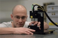
Researchers at the National Physical Laboratory (NPL) have developed a new means to ensure that a screening technique using Optical Coherence Tomography (OCT) systems can be relied upon by clinicians so that they can accurately spot early signs of cancer.
OCT is an increasingly popular method for looking beneath the surface of certain materials, notably human tissue. Although it can only be used to image tissue at depths of a few millimetres, it can produce higher-resolution images than either MRI or ultrasound, making it suitable for detecting changes in tissue structure that can indicate the early stages of cancer.
Indeed, OCT systems such as those from Orpington Kent-based Michelson Diagnostics have already proven capable of imaging microstructures in living tissue in real time at depths of 1mm or more at a resolution of less than 10µm.
In a simple OCT system, a source of light from a laser is separated into two beams by a Michelson interferometer — one beam is focused on a sample through a lens and the other half is directed towards a reference mirror. The light reflected from the mirror and from the sample is then sent to a detector that measures the degree of interference between the two sources. The signal from the detector is then processed and used to create a spatial reflectivity map of the sample that contains both depth and intensity information.
According to Dr Pete Tomlins, a senior research scientist in the Optical Technologies and Scientific Computing Division at the NPL, the depth and lateral resolution of such OCT systems are constrained by the physical nature of the optical pathways employed in the system.
Because of such constraints, OCT instrument manufacturers need to be able to align the optical pathways in the machines to optimise their performance, as well as to ensure that the systems will operate within specification once they are deployed in the field. Now, a new product developed at the NPL — called a ‘point-spread phantom’ — will allow them to do just that.
OCT is increasing in popularity as a method of looking underneath the surface of human tissue
The phantoms themselves are translucent cylinders of resin containing specially arranged submicron spherical metal particles designed to reflect light in a specific way. By viewing the images of the particles in the phantom with an OCT machine and then analysing the image with NPL software, users can be certain the machine is producing precise and reliable images.
In use, a phantom is first placed under the imaging optics of an OCT instrument, which then scans the entire volume, creating a three-dimensional image volume. From that volume, a threshold algorithm then detects the position of each of the metal particles in the phantom.
Because the lenses in OCT systems are not perfect, when light reflected from a submicron phantom dot travels through them, the image captured is not the same as the original. The lens introduces a small amount of blur — the light is spread out in what is known as a point-spread function.
Once the software has located all of the points, it then fits a Gaussian mathematical function to the data that has been collected, which provides a representation of the point-spread function of each the images of the spherical metal particles. The data can be further interpreted to determine the lateral and depth resolution of each of the points, as well as the lateral resolution of the instrument across the entire plane scanned.
‘From that data, a manufacturer could identify where the useable field of view of the instrument is — an incredibly important detail to know,’ said Tomlins.
By determining the resolution of each of the points in the sample, it is also possible to create histograms from the data, which from a statistical perspective would enable a manufacturer to determine the average performance of an instrument. A further analysis would allow a manufacturer to examine which parts of an optical system might be creating specific aberrations and where it could be optimised.
Michelson Diagnostics is the first UK company to use NPL’s phantoms to validate the accuracy of its machines and to optimise its performance. Chief executive Jon Holmes said: ‘NPL’s phantoms and analysis have enabled us to validate our performance claims beyond doubt. We expect this validation to give OCT technology the backing it needs to become standard in hospitals around the world and thereby make an important leap in the battle against cancer.’
The system developed at Michelson Diagnostic is claimed to provide Optical Coherence Tomography (OCT) images at a lateral resolution at least double that of competitor instruments because it uses four beams to scan the sample.This provides a user with much higher detail of the sub-surface tissue than would otherwise be possible.
Aside from using the phantoms to validate the specification of the machine, the engineers at Michelson Diagnostics also deployed them to provide a visual confirmation that all the optical channels in the instrument were aligned to give the system the best visual performance.
Tomlins claims that there has also been interest in this technique from many OCT companies. ‘Now we have proven that the phantom technology works, we would like to beta test it with many organisations — a testing programme would give us more feedback on how the system could potentially be used,’ he added.
Researchers at the US Food and Drug Administration (FDA) have been in contact with the NPL team too. They have been working on developing a similar phantom for the US market that is based on a flexible silicone, rather than a resin. Both organisations are currently working together to develop a standard test methodology for OCT systems using phantoms that can characterise the instruments before clinical use and can track instrument performance once in the clinic.
We expect our validation of NPL’s phantoms to give OCT the backing to become standard in all hospitals
Jon Holmes, Michelson Diagnostics
‘We anticipate that eventually such phantoms will be shipped with OCT systems when they are used clinically, together with a set of guidelines highlighting how they are to be used,’ said Tomlins.

While the phantoms can be used to spatially map the resolution of the instrument for alignment and verification purposes, the NPL researchers are now aiming to use the point-spread data collected from the phantoms to help develop a universal means to deconvolve the images produced by all OCT machines. This will allow them to remove the aberration of the point-spread function from the image data and improve the resolution throughout the imaging volume of an instrument.
‘By removing the point-spread function from the data, we have actually seen an improvement of several microns in resolution of an OCT instrument, which is very exciting. We have demonstrated that you can now resolve two points that previously couldn’t be resolved,’ said Tomlins.
The deconvolution technique will also allow the data collected from any OCT machine to be quantitatively analysed independent of any artefacts that may have been introduced by the instrument itself. In doing so, the instrument manufacturers will then be able to offer their customers an objective, rather than a subjective, way to classify pre-cancerous tissue. ‘If you can remove the effect of the point-spread function created by the systems in a standard way, you can make objective measurements of human issue and objective classifications of pre cancer,’ added Tomlins.
He feels that while OCT is now a very powerful imaging technology, it will truly realise its potential in a clinical situation when it can provide a clinician with this quantitative means of evaluating a tissue sample. NPL’s work developing the phantoms has made an important contribution to bringing the day when that happens a lot closer.
Key facts
- OCT systems need to be more reliable to spot cancer at an early stage
- Viewing images using a point-spread phantom increases accuracy
- A standard test method for systems using phantoms is being developed
- OCT will realise its potential quantitatively evaluating tissue samples


Swiss geoengineering start-up targets methane removal
No mention whatsoever about the effect of increased methane levels/iron chloride in the ocean on the pH and chemical properties of the ocean - are we...