
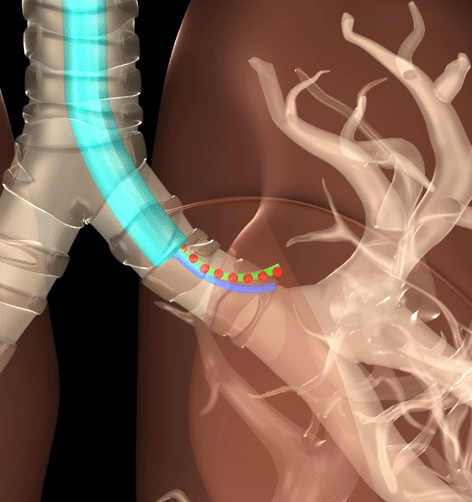
Many surgical procedures now use long, thin devices – such as ‘steerable needles’ – that can be inserted into a patient’s body through a small incision and then steered to a target location.
These minimally invasive procedures - including laparoscopic or other procedures such as biopsis or drug-delivery with steerable needles - allow doctors to perform surgeries without having to make major incisions, which decreases the risk of infection and shortens the patient’s recovery time.
However, these techniques pose a challenge to surgeons, because it is difficult for them to determine precisely where the surgical device is in the patient’s body.
One solution to the problem is to use X-rays to track the progress of the surgical device in the patient. But doctors want to minimize the number of X-rays taken, in order to limit the patient’s exposure to radiation.
‘We have now developed an algorithm to determine the fewest number of X-rays that need to be taken, as well as what angles they need to be taken from, in order to give surgeons the information they need on a surgical device’s location in the body,’ said Dr. Edgar Lobaton, an assistant professor of electrical and computer engineering at North Carolina State University and lead author of a paper on the research carried out with the University of North Carolina at Chapel Hill (UNC).
The new tool is a computer program that allows surgeons to enter what type of procedure they’ll be performing and how precise they need the location data to be. Those variables are then plugged into the algorithm developed by the research team, which tells the surgeon how many X-rays will be needed – and from which angles – to produce the necessary location details.
According to a statement, if a surgeon needs only a fairly general idea of where a device is located, only two or three X-rays may be needed – whereas more X-rays would be required if the surgeon needs extremely precise location data.
The paper, ‘Continuous Shape Estimation of Continuum Robots Using X-ray Images,’ will be presented at the IEEE International Conference on Robotics and Automation, being held in Karlsruhe, Germany, May 6-10.
The paper was co-authored by Jingua Fu, a former graduate student at UNC; Luis Torres, a Ph.D. student at UNC; and Dr. Ron Alterovitz, an assistant professor of computer science at UNC. The research was supported by the National Science Foundation and the National Institutes of Health.


April 1886: the Brunkebergs tunnel
First ever example of a ground source heat pump?