Called GLIM (gradient light interference microscopy), the method is claimed to solve the challenge of imaging thick, multicellular samples.
The new technique, published in Nature Communications, brought together electrical and computer engineering professor Gabriel Popescu and animal sciences professor Matthew Wheeler in a collaborative project through the Beckman Institute for Advanced Science and Technology at the University of Illinois (UI).
Some forms of biomedical microscopy require light to be shined through thin slices of tissue to produce an image, whereas others use chemical or physical markers that allow the operator to find specific objects within a thick sample, but those markers can be toxic to living tissue, Popescu said.
"When looking at thick samples with other methods, your image becomes washed out due to the light bouncing off of all surfaces in the sample," said graduate student Mikhail Kandel, the co-lead author of the study. "It is like looking into a cloud."
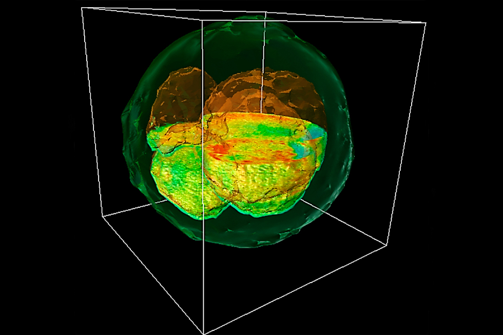
According to UI, GLIM can probe deep into thick samples by controlling the path length over which light travels through the specimen. The technique allows the researchers to produce images from multiple depths that are then composited into a single 3D image.
To demonstrate the new method, Popescu's group collaborated with Wheeler and his team to examine cow embryos.
"One of the holy grails of embryology is finding a way to determine which embryos are most viable," Wheeler said. "Having a non-invasive way to correlate to embryo viability is key; before GLIM, we were taking more of an educated guess."
Those educated guesses are made by examining factors like the colour of fluids inside the embryonic cells and the timing of development, among others, but there is no universal marker for determining embryo health, Wheeler said.
"This method lets us see the whole picture, like a three-dimensional model of the entire embryo at one time," said Tan Nguyen, the other co-lead author of the study.
Choosing the healthiest embryo is not the end of the story, though. "The ultimate test will be to prove that we have picked a healthy embryo and that it has gone on to develop a live calf," said Marcello Rubessa, a postdoctoral researcher and co-author of the study.
The team hopes to apply GLIM technology to human fertility research and treatment, as well as a range of different types of tissue research.


Swiss geoengineering start-up targets methane removal
No mention whatsoever about the effect of increased methane levels/iron chloride in the ocean on the pH and chemical properties of the ocean - are we...