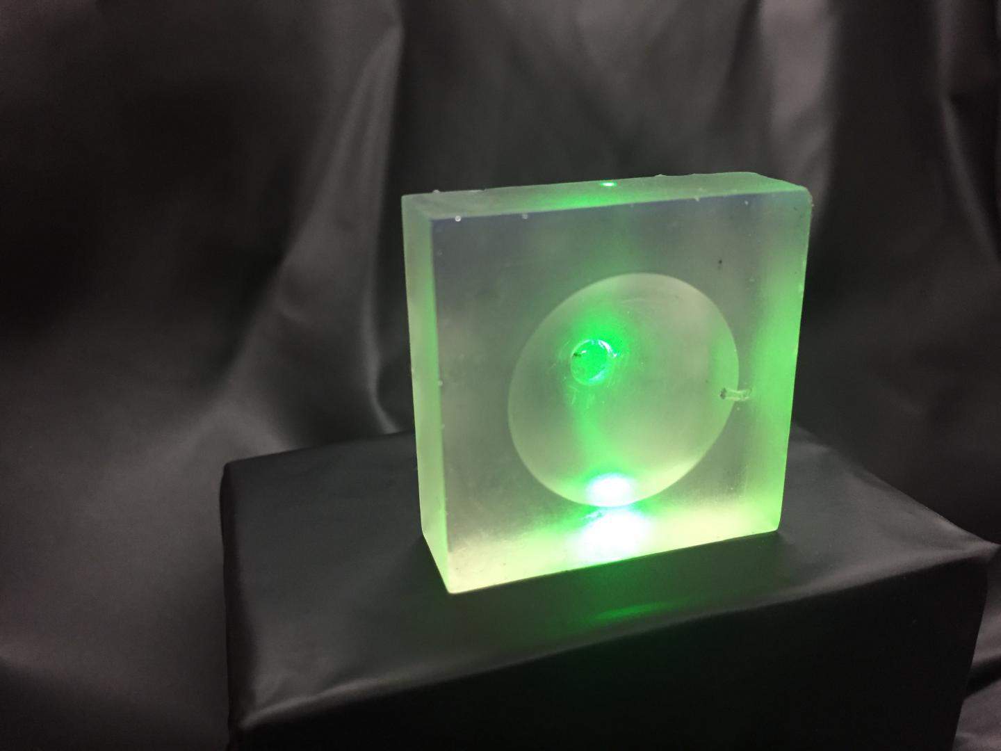
The abilities of ultrasound waves in medicine can be enhanced by directing them through 3D printed lenses, according to researchers at Nanyang Technological University in Singapore. The lenses, which are made of resin, allow the waves to be brought to a much sharper focus than is possible with conventional glass lenses, which produces better images and will allow clinicians to work with greater control and precision.
Ultrasound beams are produced by firing high-frequency sound waves at a lens, which focuses the waves. Conventionally, these lenses are made of glass and are cylindrical or spherical. Because of these relatively simple geometries, the lenses cannot focus the beams tightly onto a target.
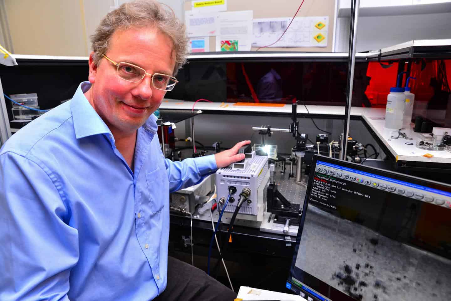
Using 3D printing to make resin lenses allows much more complex geometry to be incorporated into their shapes, which means the ultrasound can be focused onto multiple sites or shaped to direct energy in a particular way. Associate Prof Claus-Dieter Ohl of Nanyang’s School of physical and mathematical sciences, who led the multidisciplinary team behind the research, said: "3D printing reinvents the manufacturing process, enabling the creation of unique and complex devices. In turn, the way medical devices are created needs to be rethought. This is an exciting discovery for the scientific community as it opens new doors for research and medical surgery.”
The lenses are not only more versatile than those made of glass, but are cheaper and easier to produce. As well as being used to image inside the body, they could also be used in ex-vivo research, Ohl suggests; for example, to measure the elastic properties of cancerous and healthy cells growing in a Petri dish: this could be used to help distinguish tumours from healthy tissue in subsequent scans. “In most medical surgeries, precision and non-invasive diagnosis methods are crucial,” Ohl said. “This novel device not only determines the focus of the wave but also its shape, granting greater accuracy and control to medical practitioners.”
The lenses could also be used to enhance the therapeutic use of ultrasound, for example in breaking up blood clots, bursting tumour cells or controlled release of drugs from an implant. The Nanyang team describes this research in a paper in the journal Applied Physics Letters.


Red Bull makes hydrogen fuel cell play with AVL
Formula 1 is an anachronistic anomaly where its only cutting edge is in engine development. The rules prohibit any real innovation and there would be...