
The researchers said they have clinically validated and tested the AI on large sets of CT scans, and found that it could detect, segment, quantify and differentiate different types of brain lesions.
MORE MEDICAL CONTENT FROM THE ENGINEER
The results could be useful in large-scale research studies, for developing more personalised treatments for head injuries and, with further validation, could be useful in certain clinical scenarios, such as resource-limited areas with few radiologists.
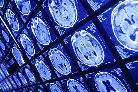
Head injury affects up to 60 million people each year and is the leading cause of mortality in young adults. When a patient has had a head injury, they are often sent for a CT scan to check for blood in or around the brain, and to help determine whether surgery is required. However, of all the patients who have a head injury, between 10 and 15 per cent have a lesion that can be seen on a CT scan.
"CT is an incredibly important diagnostic tool, but it's rarely used quantitatively," said co-senior author Professor David Menon, from Cambridge's Department of Medicine. "Often, much of the rich information available in a CT scan is missed, and as researchers, we know that the type, volume and location of a lesion on the brain are important to patient outcomes."
According to Cambridge University, different types of blood in or around the brain can lead to different patient outcomes, and radiologists will often make estimates in order to determine the best course of treatment.
"Detailed assessment of a CT scan with annotations can take hours, especially in patients with more severe injuries," said co-first author Dr Virginia Newcombe, also from Cambridge's Department of Medicine. "We wanted to design and develop a tool that could automatically identify and quantify the different types of brain lesions so that we could use it in research and explore its possible use in a hospital setting."
The researchers’ machine learning tool is based on an artificial neural network. They trained the tool on over 600 different CT scans, showing brain lesions of different sizes and types. They then validated the tool on an existing large dataset of CT scans.
The AI was able to classify individual parts of each image and tell whether it was normal or not. This could be useful for future studies in how head injuries progress because AI may be more consistent than a human at detecting subtle changes over time.
"This tool will allow us to answer research questions we couldn't answer before," Newcombe said in a statement. "We want to use it on large datasets to understand how much imaging can tell us about the prognosis of patients."
"We hope it will help us identify which lesions get larger and progress, and understand why they progress, so that we can develop more personalised treatment for patients in future," said Menon.
The team’s findings are reported in The Lancet Digital Health.


Nanogenerator consumes CO2 to generate electricity
Whoopee, they've solved how to keep a light on but not a lot else.