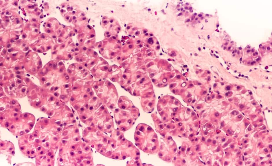
Photoacoustic tomography is a non-invasive technique that converts absorbed optical energy into acoustic signal. Pulsed light is sent into body tissue, creating a small increase in temperature that expands tissue to an acoustic response detected by an ultrasound transducer. Ultrasound data is then used to visualise the tissue.
“The nice thing about photoacoustic tomography is the compositional information,” said Craig Goergen, an assistant professor in Purdue University’s Weldon School of Biomedical Engineering. “It provides information about where blood and lipid are located, along with other essential information.”
The results of a study describing an adjustable photoacoustic probe with improved light delivery and image quality have been published in Photoacoustics.
According to Purdue, the system provides real-time compositional information of body tissue without the need for contrast agents and with better depth penetration compared with conventional optical techniques.
Photoacoustic tomography can be used to detect or monitor diseases including cardiovascular disease, diabetes, and cancer. These conditions are estimated by the US Centers for Disease Control and Prevention to cost over $718bn a year.
“That means there will be a great need for medical imaging. Trying to diagnose these diseases at an earlier time can lead to improved patient care,” Goergen said. “We are in the process now of trying to use this enhanced imaging approach to a variety of different applications to see what it can be used for.”
Among other potential uses for photoacoustic tomography is the mapping of lipid deposition within an arterial wall that can cause other health problems, measuring cardiac tissue damage and tumour biopsies. Using photoacoustic tomography for intraoperative tumour biopsies could help surgeons make sure they remove all traces of cancer from a patient, Goergen said.
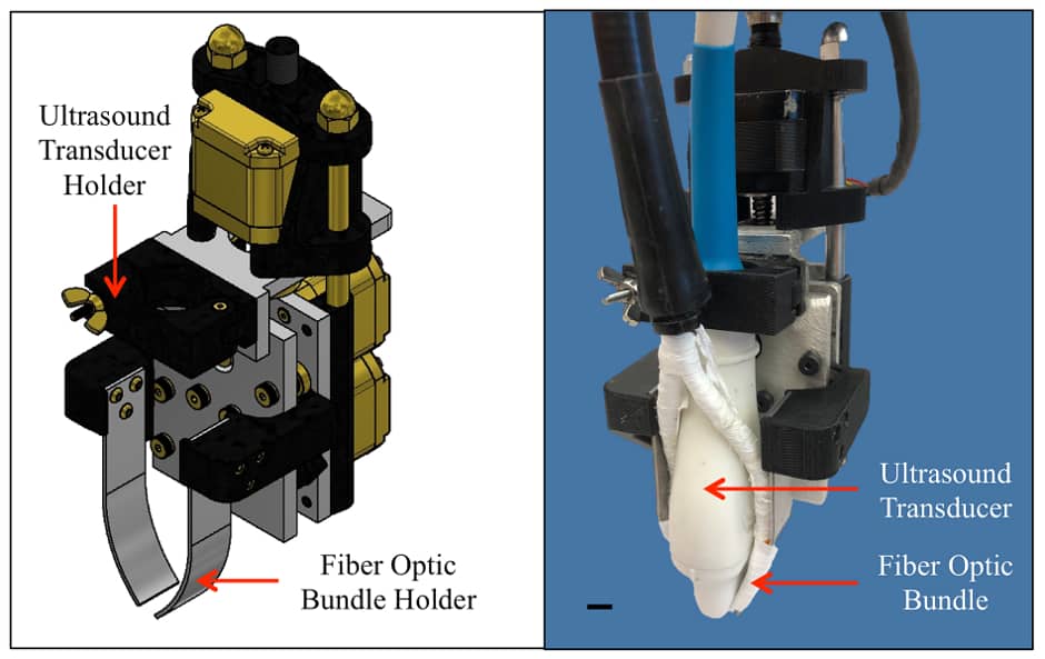
One of the challenges of photoacoustic tomography is improving the penetration depth and signal-to-noise ratio past optical absorbers. The researchers believe creating optical manipulation techniques to maximise photon density could provide a solution. Consequently, they have created a motorised photoacoustic holder that allows users to easily manoeuvre the aim of the device and tune the depth where light is focused, improving the light penetration depth and signal-to-noise ratio.
The researchers have a patent pending for the biomedical imaging technology. A video about the acoustic tomography is available at: https://bit.ly/2yJddb0


Swiss geoengineering start-up targets methane removal
No mention whatsoever about the effect of increased methane levels/iron chloride in the ocean on the pH and chemical properties of the ocean - are we...