The development from a team at KAUST in Saudi Arabia involves an automated process that prints a peptide-based hydrogel scaffold containing uniformly distributed cells. According to KAUST, the scaffolds hold their shapes well and successfully facilitate cell growth that lasts for weeks.
Bioprinting microrobot holds promise for internal tissue repairs
Ceramic-based ink enables 3D-printed bone tissue
Scientists have experimented with natural and synthetic ‘bioinks’ to print out scaffolds that hold cells in place as they grow and form a tissue with a specific shape, but cell survival remains a challenge. Natural bioinks including gelatin and collagen need to be treated with chemicals or ultraviolet light to hold their shape, which affects cell viability. The synthetic polymer hydrogels tested to date also require the use of harsh chemicals and conditions that threaten cell survival.
KAUST bioengineer Charlotte Hauser led a team to develop a bioprinting process that uses ultrashort peptides as the basis of the scaffolding ink. They designed three peptides using different combinations of the amino acids isoleucine, lysine, phenylalanine and cyclohexylalanine.
The team used a novel triple-inlet nozzle to do the printing with the peptide bioink entering one inlet, a buffer solution into another, and cells added through a third. This allows the peptide ink to gradually mix with the buffer solution and then combine with the cells at the nozzle's outlet. Once the ink is ejected, it instantly solidifies, capturing the cells within its structure.
"It's challenging to find a cell-friendly biomaterial that supports long-term cell survival and is also printable," PhD student Hepi Hari Susapto said in a statement. "Our bioinks made from self-assembling ultrashort peptide hydrogels efficiently address this challenge."
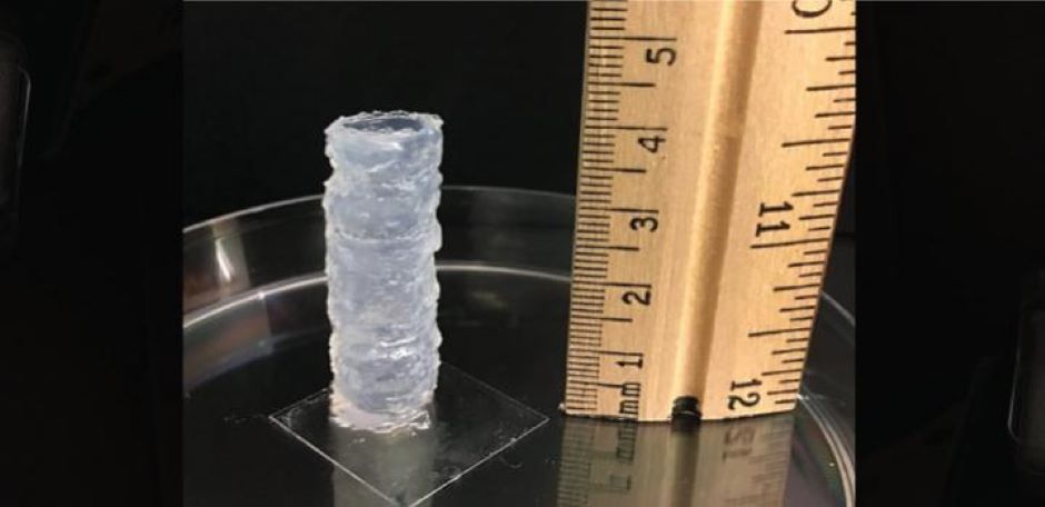
The team printed cylinders up to 4cm tall and a human-like nose, which are all said to have held their shapes.
Human fibroblasts, human bone marrow mesenchymal stem cells and mouse brain neurons reportedly survived and proliferated well within the hydrogel matrix. The scientists further induced bone marrow mesenchymal stem cells to differentiate inside a printed scaffold into elastic cartilage-like tissue within a period of four weeks.
The team is now working on changing the surface chemistry of their bioinks so that they more closely resemble the cell environment in the human body.
"Our next step is to bioprint 3D disease models and miniature organs for high-throughput drug screening and diagnosis," said Hauser. "These could help reduce the time and cost of searching for more effective and personalised drugs."
The team's findings have been published in Nano Letters.


Nanogenerator consumes CO2 to generate electricity
Whoopee, they've solved how to keep a light on but not a lot else.