A Johns Hopkins electrical engineer is building a tool to help brain surgeons locate and get a clear look at cancerous tissue.
In some cases, the device could eliminate the need to cut into the brain for a traditional biopsy, a procedure that can pose risks to the patient.
Researcher Jin U Kang said: 'The idea is to provide instant high-resolution pictures of a small segment of the brain without actually touching the tissue.
'These pictures could let the doctor conduct a “virtual biopsy” to see where the tumour is and whether it is benign or malignant.
'And when it's time to cut out the cancer, these images could help a surgeon see and avoid healthy tissue.'
Kang's concept recently received a financial boost that should help move it from the drawing board to the operating room.
He was awarded $450,000 (£271,629) in US federal stimulus package funds to help develop the surgical instrument further.
The two-year grant was provided by the
Kang’s team has made great strides in refining the technology, but the surgical tool has not yet been tried out on human patients.
The federal grant will enable the researchers to begin animal and human cadaver testing in the coming months.
Human patient trials could begin within five years.
To give doctors this detailed view of brain tissue, Kang’s device employs a thin optical fibre to direct low-powered laser light onto the area the surgeon wants to examine.
When the light strikes the tissue, most of it bounces away in a scattered, incoherent manner.
But a small portion of light is scattered back, collected and used to construct a high-resolution three-dimensional picture of the tissue, down to the cellular level.
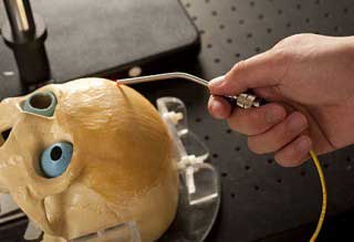
In this demonstration using a model skull, Jin Kang’s device employs thin optical fibre to direct harmless low-powered laser light onto the area the surgeon wants to examine
The images produced by the optical coherence tomography system are significantly sharper than those produced by MRI or ultrasound equipment, Kang said, and should give surgeons a better look at the boundaries of a tumour and the presence of blood vessels and healthy tissue that must be preserved.
Yet compared with the older, widely used imaging systems, the new system is expected to be much less expensive, perhaps less than $10,000.


Project to investigate hybrid approach to titanium manufacturing
What is this a hybrid of? Superplastic forming tends to be performed slowly as otherwise the behaviour is the hot creep that typifies hot...