Surgeons operating on cancer patients have no way of determining in real-time whether they have removed all of the diseased tissue, which is the key to successful surgery.
However, that could soon be a thing of the past.
Researchers in
The technique shows particular promise for improving surgery on breast, prostate and lung cancer, the tumour boundaries for which can be difficult to track at advanced stages.
The technique can also help cancer surgeons avoid cutting critical structures such as blood vessels and nerves.
Dr John Frangioni, project director of Beth Israel Deaconess Medical Center (BIDMC) in Boston and co-director of its Center for Imaging Technology and Molecular Diagnostics, said: 'This technique is really the first time that cancer surgeons can see structures that are otherwise invisible, providing true image-guided surgery. If we are able to see cancer, we have a chance of curing it.'
The system is called FLARE, or Fluorescence-Assisted Resection and Exploration.
Under development for the past decade, the portable system consists of a near-infrared (NIR) imaging system, a video monitor and a computer.
Frangioni added: 'The system has no moving parts, uses LEDs instead of lasers for excitation, makes no contact with the patient and is sterile.'
The unique system uses special chemical dyes called NIR fluorophores that target specific structures such as cancer cells when the dyes are injected into patients.
When exposed to NIR light, which is invisible to the human eye, the dyes or contrast agents light up the cancer cells and are shown on a video monitor.
Images of these 'glowing' cancer cells are then superimposed over images of the normal surgical field, allowing surgeons to easily see the cancer cells even in a background crowded by blood and other anatomical structures.
The computerised technique also gives physicians the power to control multiple viewing angles and different magnification levels through the use of a footswitch.
In preliminary studies, Frangioni and colleagues used FLARE to successfully visualise organs and body fluids of mice and map the lymph nodes of pigs, all in real-time.
The first human clinical trials, expected to begin this summer, involve mapping the lymph nodes of a small group of patients with breast cancer.
Broader clinical use of the device could occur within five years.
In the future, fluorophores could be developed to highlight nerves and blood vessels in one colour while visualising cancer cells in a different colour, allowing multiple structures to be viewed easily and even simultaneously.
Frangioni said: 'The future of the technology now is really in the chemistry. We have to develop agents for specific tumours, nerves or blood vessels we are trying to visualise.'
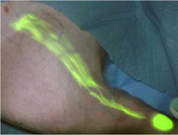
Photo of a pig's hind leg viewed after injection with near-infrared contrast agents and imaging with near-infrared light to highlight lymph flow


Red Bull makes hydrogen fuel cell play with AVL
Formula 1 is an anachronistic anomaly where its only cutting edge is in engine development. The rules prohibit any real innovation and there would be...