The compact broadband near-infrared spectroscopy (NIRS) system has been developed by UCL’s Multimodal-Spectroscopy (MMS Group) team.
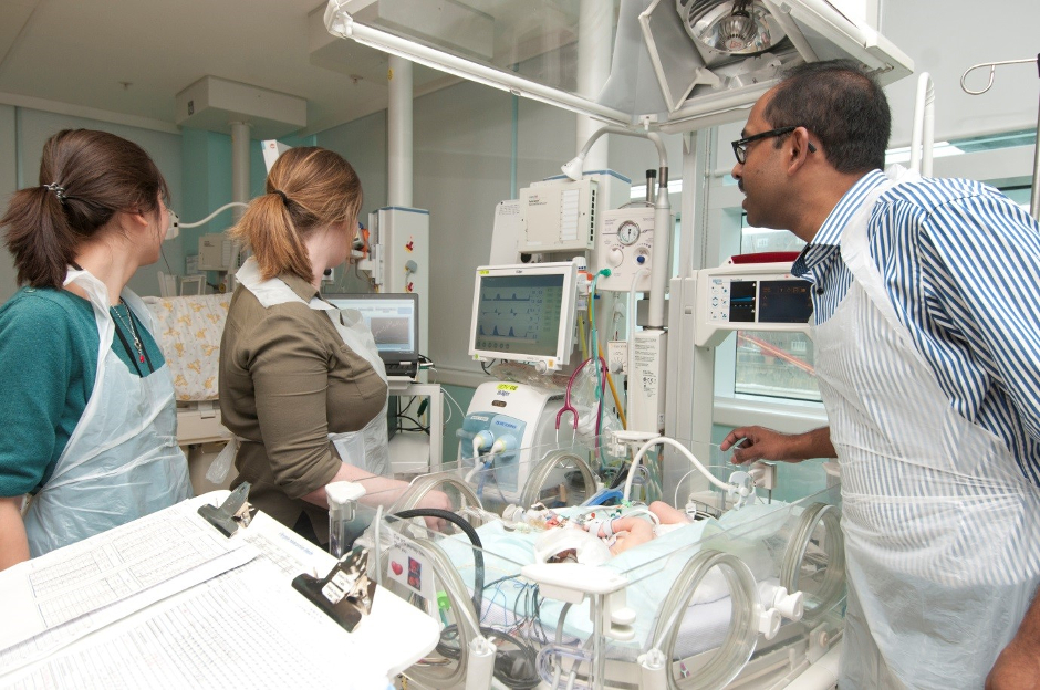
The portable device, which is small enough for use in neonatal intensive care units (NICU) and accident and emergency (A&E) rooms, is currently being used in preclinical studies and in clinical studies in the neonatal unit at University College London Hospital (UCLH).
During birth, disruptions in blood and oxygen supply to a baby’s brain can stop it from working properly. This can lead to an acute injury to the developing brain, and can ultimately lead to significant disability and death. While many babies make a partial to full recovery, some will develop cerebral palsy or behavioural problems.
Detecting and monitoring newborn brain function following such disruption is vital for doctors if they are to understand the effects of the injury and neurodevelopmental outcomes for the baby.
CYRIL is claimed to offer a safe way of shining light through the brain tissue of a newborn baby and detecting its function with a sensitive digital camera. This reveals detailed information about oxygen and metabolism levels, providing potentially life-saving information for doctors. The research team have demonstrated that the metabolic information collected by CYRIL can identify brain injury severity.
By using hundreds of wavelengths of light, broadband NIRS measurements of cytochrome-c-oxidase (CCO) can yield crucial information about brain metabolism at the cot side. CCO catalyses over 95 per cent of oxygen to produce energy, providing information about oxygen consumption at cellular level.
CLICK HERE FOR MORE MEDICAL & HEALTHCARE NEWS
CYRIL (CYtochrome-c-oxidase Research Instrument and appLication) simultaneously measures changes in brain oxygenation and haemodynamics via estimation of the changes in haemoglobin concentration; in addition to oxygen utilisation via the measurement of the oxidation state of CCO.
Team leader Dr Ilias Tachtsidis said that future work will focus on transforming these optical devices to compact clinical instruments that can be operated by medical doctors and nurses to provide real-time information on the brain function.
Dr Subhabrata Mitra, a clinical academic consultant in the neonatal unit at UCLH said: “[CYRIL] has shown the potential to become a fantastic bedside neuromonitoring tool for babies that already have or are prone to developing newborn brain injury.
“It has the ability to identify babies based on injury severity early on following hypoxic ischaemic encephalopathy (HIE), can give us an early diagnostic direction in cases of neonatal stroke and also gives as important information regarding the pathophysiological changes in brain during neonatal seizures.”
In a separate development, the MMS Group has also launched MetaboLight to share their research and encourage the next generation of medical physicists and biomedical engineers.


Red Bull makes hydrogen fuel cell play with AVL
Formula 1 is an anachronistic anomaly where its only cutting edge is in engine development. The rules prohibit any real innovation and there would be...