fibre-optic ultrasonic imaging probe developed at Nottingham University.
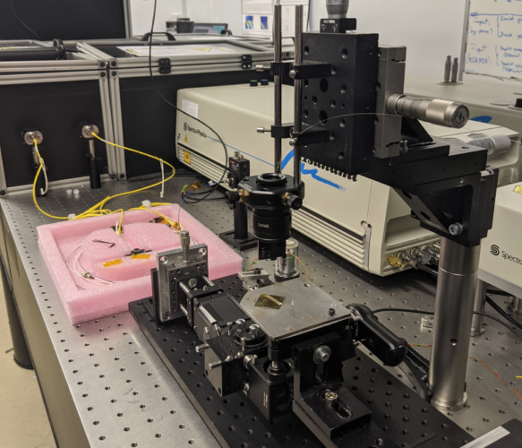
Claimed to be a world first, the ultrasonic imaging system, which can be deployed on the tip of a thin optical fibre, will be insertable into the human body to visualise cell abnormalities in 3D.
MORE FROM MEDICAL & HEALTHCARE
The new technology produces microscopic and nanoscopic resolution images likely to help clinicians to examine cells inhabiting hard-to-reach parts of the body, such as the gastrointestinal tract.
The EPSRC-funded device is said to deliver a level of performance only possible in advanced research labs. It also reduces the need for conventional fluorescent labels, which can be harmful to human cells in large doses. The findings are detailed in Light: Science & Applications.
“We believe its ability to measure the stiffness of a specimen, its bio-compatibility, and its endoscopic-potential, all while accessing the nanoscale, are what set it apart,” said paper author Dr Salvatore La Cavera III. “These features set the technology up for future measurements inside the body; towards the ultimate goal of minimally invasive point-of-care diagnostics.”
The prototype non-invasive imaging tool, described as a “phonon probe”, is capable of being inserted into a standard optical endoscope. Combining optical and phonon technologies could be advantageous because they accelerate the clinical workflow process and reduce the number of invasive test procedures for patients.
“Techniques capable of measuring if a tumour cell is stiff have been realised with laboratory microscopes, but these powerful tools are cumbersome, immobile, and unadaptable to patient-facing clinical settings. Nanoscale ultrasonic technology in an endoscopic capacity is poised to make that leap,” said Dr La Cavera, an EPSRC Doctoral Prize Fellow from Nottingham University’s Optics and Photonics Research Group.
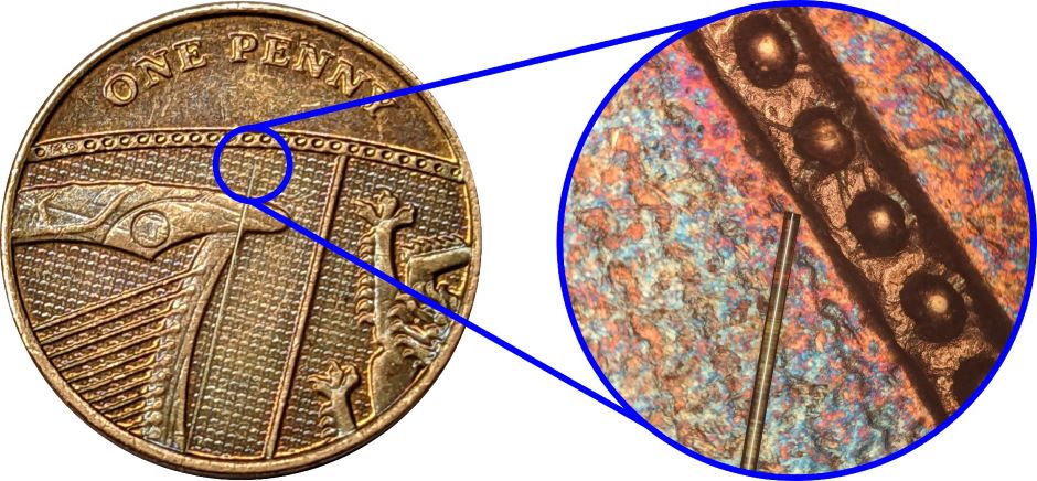
In use, the new ultrasonic imaging system uses two lasers that emit short pulses of energy to stimulate and detect vibrations in a specimen. One of the laser pulses is absorbed by a layer of metal – a nano-transducer – fabricated on the tip of the fibre; a process which results in high-frequency phonons getting pumped into the specimen. Then a second laser pulse collides with the sound waves. By detecting these “collided” laser pulses, the shape of the travelling sound wave can be recreated and displayed visually.
The detected sound wave encodes information about the stiffness of a material and even its geometry. The Nottingham team was the first to demonstrate this dual-capability using pulsed lasers and optical fibres.
In the paper, the researchers demonstrate that the technology is compatible with a single optical fibre and the 10-20,000 fibres of an imaging bundle, as used in conventional endoscopes.
Consequently, superior spatial resolution and wide fields of view could routinely be achieved by collecting stiffness and spatial information from multiple different points on a sample, without needing to move the device. According to the university, this heralds a new class of phonon endoscopes.
Beyond clinical healthcare, fields such as precision manufacturing and metrology could use this high-resolution tool for surface inspections and material characterisation. Technologies such as 3D bio-printing and tissue engineering could also use the phonon probe as an inline inspection tool by integrating it directly to the outer diameter of the print-needle.
The team will next develop a series of biological cell and tissue imaging applications in collaboration with the Nottingham Digestive Diseases Centre and the Institute of Biophysics, Imaging and Optical Science at Nottingham University. Their aim is to create a viable clinical tool in the coming years.


Glasgow trial explores AR cues for autonomous road safety
They've ploughed into a few vulnerable road users in the past. Making that less likely will make it spectacularly easy to stop the traffic for...