
Deep brain stimulation involves implanting electrodes into the brain to produce electrical impulses that control abnormal movement, but current implant materials can present anomalies when the patient requires further medical evaluation via an MRI. This is because the metal electrode may react to the magnetic fields and vibrate, generate heat or damage the brain.
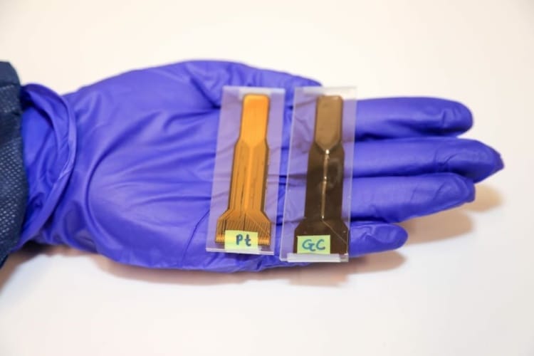
Now, a study published in Nature Microsystems & Nanoengineering describes a promising improvement to the procedure developed by San Diego State University engineers, in collaboration with researchers at Karlsruhe Institute of Technology (KIT), Germany. The SDSU research team created a glassy carbon electrode as an alternative to the metal version, and new findings show it does not react to MRI scans.
Deep brain stimulation used to slow down Alzheimer’s associated decline
Untethered electrode sheds light on therapeutic uses of neural stimulation
First developed in 2017 in researcher Sam Kassegne's MEMS lab at SDSU, the carbon version is designed to last longer in the brain without corroding or deteriorating and to emit and receive stronger signals. In 2018, the researchers showed that while the metal electrode degrades after 100 million cycles of electrical impulses applied to it, the glassy carbon material survived 3.5 billion cycles.
Until now, the electrodes have been made from thin-film platinum or iridium oxide. According to SDSU, such metal-based electrodes can produce heat, interfere with the MRI images by creating bright spots that block views of the actual area in the brain being studied, and can become magnetised and move or vibrate when patients undergo scans, causing discomfort.
"Our lab testing shows that unlike the metal electrode, the glassy carbon electrode does not get magnetised by the MRI, so it won't irritate the patient's brain," said Surabhi Nimbalkar, first author and doctoral candidate.
In addition, it can read chemical and electrical signals from the brain, while the metal-based electrodes can only read electrical signals, so the carbon material is multi-modal as well as MRI compatible.
"It's supposed to be embedded for a lifetime, but the issue is that metal electrodes degrade, so we've been looking at how to make it last a lifetime," said Kassegne, senior author and professor of mechanical engineering at SDSU. "Inherently, the carbon thin-film material is homogenous…so it has very few defective surfaces. Platinum has grains of metal which become the weak spots vulnerable to corrosion."
Collaborators at KIT developed a novel instrument which enables precise measurements of vibrations during MRI. Working with the SDSU team, they were able to test the novel carbon electrodes directly in the MRI scanner, and confirm it was a safer, better alternative.
With lab testing completed, Kassegne's collaborators on the clinical side will now test the carbon electrode in patients, while Nimbalkar and Kassegne work on testing different forms of carbon to be used in future electrodes.


Swiss geoengineering start-up targets methane removal
No mention whatsoever about the effect of increased methane levels/iron chloride in the ocean on the pH and chemical properties of the ocean - are we...