Understanding the workings of the brain has been a major goal for medicine for centuries. Insights into how brains send signals between their many structures would give doctors valuable insights into the many different types of brain disorders, and allow them to form links between psychological conditions and physical processes, as well as into brain disorders like epilepsy and dementia.
The ability to implant electrodes into the brain to detect signals has allowed much data to be developed, but a new invention from Swedish researchers at Lund University could considerably enhance the capabilities of the technique. The team has developed soft electrodes that are much more compatible with living brain tissue than the stiff, inflexible electrodes that have been the only option up to now.
Brain electrodes are currently either completely rigid, or if they are flexible at all the contain solid chips which create stiff sections. When implanted into the brain — which floats in liquid inside the skull and moves around whenever the head moves and even with breathing — the electrodes rub against brain tissue and damage or even kill the adjacent cells; which are precisely the structures from which they are supposed to be gathering data. This is a particular problem for studies which take place over an extended time period.
“The signals then become misleading or completely non-existent. Our new technology enables us to implant as flexible electrodes as we want, and retain the exact shape of the electrode within the brain”, says Johan Agorelius, a doctoral student working with research directors Professor Jens Schouenborg and Lina Pettersson.
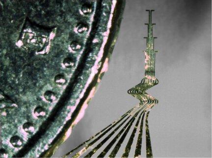
The team has developed electrodes cut by laser from a 10µm-thick sheet of gold, forming leads which are very flexible in three dimensions. They insulated the leads in parylene C, a very soft biocompatible polymer; formed the electrodes into the correct shape to monitor particular structures, then ‘locked’ this shape by encapsulating the electrode in a stiff gelatine shell.
When implanted, the shell dissolves harmlessly, leaving the biocompatible electrode free to adjust its form with brain movement and gather data without damaging the tissue.
“This technology retains the electrodes in their original form inside the brain and can monitor what happens inside virtually undisturbed and normally functioning brain tissue”, said Agorelius. “This creates entirely new conditions for our understanding of what happens inside the brain and for the development of more effective treatments for diseases such as Parkinson’s disease and chronic pain conditions than can be achieved using today’s techniques”, Schouenborg added.
The team describes in the journal Frontiers in Neuroscience how they made the electrodes, and details trials on rats, as explained further in the video below.


Swiss geoengineering start-up targets methane removal
No mention whatsoever about the effect of increased methane levels/iron chloride in the ocean on the pH and chemical properties of the ocean - are we...