This is detailed enough to spot the first signs of tumours in specific cells and is about 100 times higher resolution than X-Ray, computed tomography (CT) and Magnetic Resonance Imaging (MRI) machines can provide.
Low-cost OCT scanner promises to save eyesight
The technology behind the device is a result of six years of optical imaging research and was jointly developed by a team from Nanyang Technological University, Singapore (NTU Singapore) with researchers at the Harvard Medical School and the University of Alabama.
Using micro Optical Coherence Tomography (OCT), the device emits a spectrum of light between 700 to 950nm (near-infrared light) which penetrates human tissue and organs. The device then measures the delay time of the ‘echo’ from its light waves as they strike different tissue structures. This information will then be used to construct cross-section images of what is being scanned.
The results are sent in real-time to a computer system running software developed at NTU, which assists in diagnosis by assembling the 2D cross-section images into a three-dimensional picture and rendering different parts in colour.
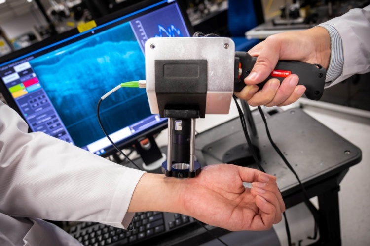
The NTU researchers said their prototype was designed to be used by medical professionals who do not specialise in imaging or pathology, allowing them to scan patients using the new device in clinics or at the bedside, negating the need for an MRI or CT scan at a specialised facility.
In a statement, NTU Associate Professor Liu Linbo, said, “Our device is a fraction of the size of existing machines and produces clear, high-resolution images in real-time. It uses light to harmlessly penetrate the skin, and it does not involve specialised lead-shielded X-ray equipment or MRI scanners. It is small enough to be handheld, so images could be captured by the bedside.”
The prototype device has undergone clinical trials at Wuhan University’s Endoscopic Centre and has reportedly shown promise in detecting abnormal colon polyps to the same level of accuracy as trained pathologists.
During the preliminary trial at the Renmin Hospital of Wuhan University, endoscopists used the device on 58 tissue samples from patients known to have colon polyps – abnormal growths in the colon or rectum. The samples were imaged in real-time by the device, and its assessment of whether they were malignant or benign was found to be 95 per cent accurate after comparison with an evaluation of the same samples, by senior pathologists. These findings were published in Clinical and Translational Gastroenterology in June 2019.
The device is now being commercialised by Suzhou Sai Luo Er Medical Imaging Technology in China.


Glasgow trial explores AR cues for autonomous road safety
They've ploughed into a few vulnerable road users in the past. Making that less likely will make it spectacularly easy to stop the traffic for...