Researchers have created a handheld probe that can image individual photoreceptors in the eyes of infants, a development that could help track early brain development in children.
Based on adaptive optics, the technology from Duke University in North Carolina is expected to make it easier for doctors and scientists to observe these cells to diagnose eye diseases and make early detection of brain-related diseases and trauma.
Photoreceptors are specialised neurons comprising the light-sensing cells of the retina, an extension of the central nervous system located at the back of the eye. The retina sends signals to the brain via the optic nerve, which then processes the visual information. Studies have shown that neurodegenerative diseases, including Alzheimer's and Parkinson's, plus traumatic brain injuries, such as concussions, can alter the neuronal structures in the retina.
To study these neuronal structures, researchers commonly use an adaptive optics scanning laser ophthalmoscope (AOSLO), a non-invasive tool that provides higher image resolution than MRI.
AOSLO lets researchers visualise individual photoreceptor cells, but such systems are large, costly and complex and its use has been limited to patients who are able to sit upright and fix their gaze for several minutes, limiting its use for young children or for adults with cognitive or mobility issues.
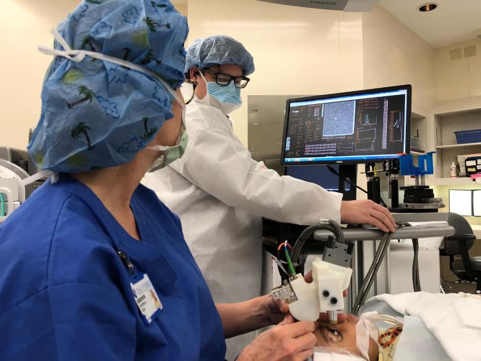
The new HAOSLO (handheld AOSLO) system measures 4 x 2 x 5.5 inches and weighs less than half a pound.
The Duke researchers are said to have transformed the AOSLO into a handheld probe by improving its optical, signal processing and mechanical designs. This included development of a new algorithm that replaces the traditional AOSLO's large wavefront sensing system, an optical component that can detect light distortion caused by the eye. The study appears in Optica.
"Other researchers have shown that the wavefront sensor can be replaced by an algorithm, but previous algorithms haven't been fast or robust enough to be used in a handheld device," said first author Theodore DuBose, a PhD student in the department of biomedical engineering at Duke. "The algorithm we developed is much faster than previous techniques and just as accurate."
According to Duke, the tool was tested in a clinical trial with 12 adults and two children, where the team reportedly demonstrated its ability to capture detailed images of photoreceptors close to the fovea, which is the centre of the retina where photoreceptors are smallest and vision is most acute.
"Our new tool is fast and lightweight so physicians can take it directly to their patients, and the probe allows us to collect images quickly, even if there is movement," said Sina Farsiu, an associate professor in the departments of biomedical engineering and ophthalmology. "These capabilities allow us to open up the pool of patients who could benefit from this technology."
The tool's new design is said to make it useful for imaging the eyes of young children. Researchers can use the tool to track early brain development in children by imaging their retina, which develops along with the central nervous system.
In a related application, surgeons will be able to see the photoreceptors at the highest resolution possible during surgery, even when a patient is under anaesthesia and in a reclined position. It could also help doctors rapidly assess possible brain trauma in athletes.
Before the researchers prepare for large-scale clinical trials, they plan to incorporate additional imaging modes for detecting other diseases.
They also have made the mechanical designs, computational algorithms and control software available online so that other scientists can adapt the new system for their scientific applications.


Nanogenerator consumes CO2 to generate electricity
Whoopee, they've solved how to keep a light on but not a lot else.