To perform intricate heart operations in babies with congenital heart disease, or to conduct complex heart repairs in adults without opening the chest, surgeons need a fast real-time imaging system that allows them to see depth.
Now they might soon have just that, thanks to a new stereoscopic system developed by Dr Robert Howe of Harvard University.
Here's how the system works: first, volumetric data from specific areas of interest in the heart are streamed in real time from an ultrasound system to a graphics workstation, which renders left-eye and right-eye views by alternating the position and orientation of the image.
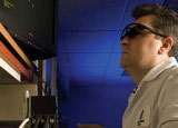
Then, the rendered volumes, immediately displayed on a conventional monitor, are synchronised with flickering shutter glasses worn by the surgeon, yielding stereo-rendered 3D ultrasound images.
The images are said to provide surgeons with significantly better spatial information and depth perception for making repairs inside the beating heart than conventional techniques.
'You definitely have depth perception,' said Dr Nikolay Vasilyev from Children's Hospital Boston. 'You feel like you're inside the heart chamber.'
Dr Vasilyev tested the glasses while operating on pigs with an atrial septal defect, a common form of congenital heart disease in which there is a hole in the wall dividing the heart's upper chambers.
Dr Vasilyev closed each defect using a catheter carrying a tiny patch, threaded into the heart through a vein. Using another device, he fastened the patch around the hole with tiny anchors. In all, he placed 64 anchors: 32 under standard 3D ultrasound guidance, and 32 using the stereoscopic vision display.
Using the stereoscopic display, Dr Vasilyev was able to place the anchors 44 per cent faster than with the standard display (9.7 versus 17.2 seconds). The tip of the anchoring device also navigated more accurately - deviation from the ideal path averaged 3.8mm, as compared with 6.1mm, a 38 per cent improvement.
The accuracy of anchor placement did not differ significantly between the two sets of tests, perhaps because of Dr Vasilyev's high level of experience and the availability of tactile information to help guide the final step of driving in the anchors. However, the speed of the anchor placement improved significantly.
Volumetric data on an atrial septal defect (arrowheads) are streamed in real time from an ultrasound system to a graphics station computer, which renders left-eye and right-eye views
Dr Pedro del Nido, chief of cardiac surgery at Children's, believes that stereoscopic imaging - coupled with recent advances in catheter-based surgical tools - will eventually allow surgeons to do much more complex operations on beating hearts, such as closing more complicated holes, shaving away excess tissue or repairing fast-moving structures like mitral or aortic valve leaflets.
Clinical trials of beating-heart surgery with the patching system could begin in children with atrial septal defects this year, Dr Vasilyev said.


Swiss geoengineering start-up targets methane removal
No mention whatsoever about the effect of increased methane levels/iron chloride in the ocean on the pH and chemical properties of the ocean - are we...