Placing and operating cameras inside living organisms is a major goal of engineers working with surgeons. However, there is an obvious problem: such devices tend to be bulky and therefore pose a risk of damaging the tissues they are imaging and passing through to reach their targets. Miniaturising endoscopes inevitably compromises the image quality they produce.
But the Johns Hopkins team, led by electrical and computer engineer Mark Foster, claims they have avoided this problem by constructing a lens-free endoscope the width of a few human hairs, capable of examining neurons firing in the brains of animals as small as mice and rats, without causing damage.
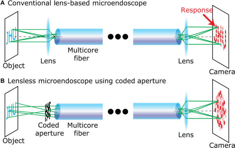
The minimum size for an endoscope with a lens is around half a millimetre. Lens-free devices, based on optical fibres, scan an area pixel by pixel, but previous versions have tended to bend in use and lose their image quality. Foster’s team tipped their device with a coded aperture, a flat grid that randomly blocks light by creating a projection in a known pattern. This is akin to randomly poking a piece of aluminium foil and letting light through all of the small holes. Although this technique creates a messy image, that image provides rich information about where the light originates, and that information can be computationally reconstructed into a clearer image.
____________________________________________________________________
Further reading
- Flat lens-free microscope could work inside body as an endoscope
- Tadpole endoscope offers new hope for gastrointestinal cancer detection
- Low cost endoscope could transform developing world cancer diagnosis
____________________________________________________________________
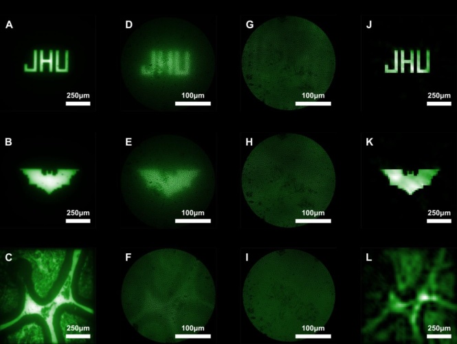
In their experiments, described in Science Advances, Foster's team looked at beads in different patterns on a slide and obtained sharp images. Moreover, the device does not need movement to focus on objects at different depths; instead, computational refocusing can determine where light originates in three dimensions. Therefore, unlike conventional endoscopes, the device does not need to be moved around to focus.


Swiss geoengineering start-up targets methane removal
No mention whatsoever about the effect of increased methane levels/iron chloride in the ocean on the pH and chemical properties of the ocean - are we...