A novel approach to 3-D printing has helped nanoengineers at the University of California San Diego to produce lifelike, functional networks of synthetic blood vessels that, in animal trials, have successfully integrated into living subjects. The team believes that this could be a first step towards a long-desired goal of being able to print functional organs and other regenerative therapies.
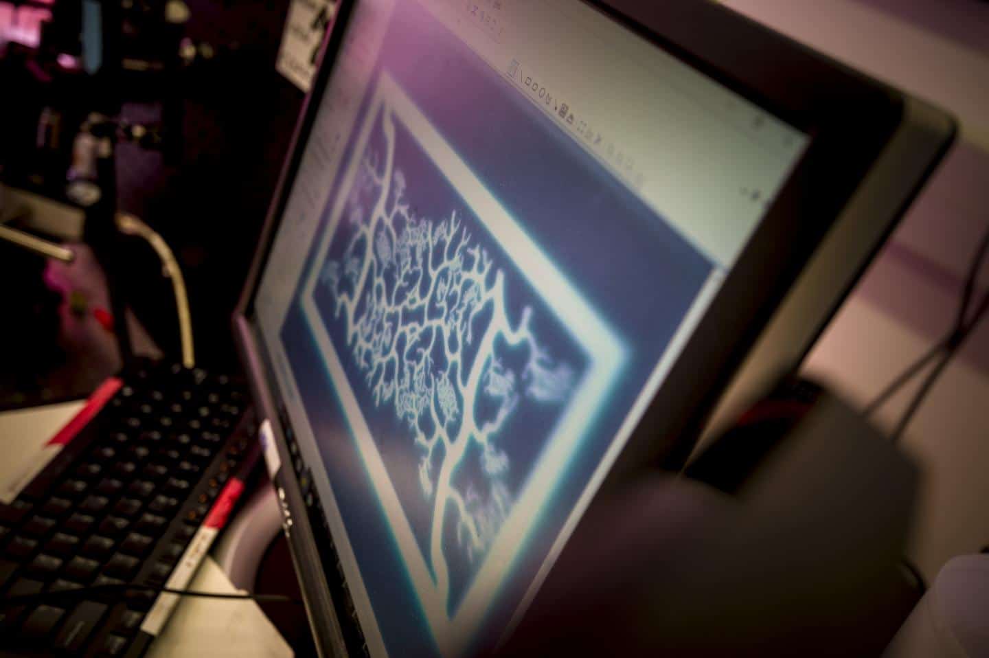
Previous work on printing blood vessels has tended to focus on printing very simple structures, such as a single vessel – effectively a tube. The San Diego research, in the Nanobiomaterials, Bioprinting, and Tissue Engineering Laboratory directed by Prof Shaochen Chen, has resulted in a network featuring branching vessels that become successively smaller, similar to those found in living tissue where they supply oxygen and nutrients and remove waste from cells.
The technique works by creating a 3D computer model of the desired structure. The computer then transfers two-dimensional snapshots of this structure from different angles to a series of motorised mirrors, which project ultraviolet light following the shape of these snapshots until onto a solution containing live cells and UV-sensitive polymers. The polymer solidifies layer by layer, trapping the cells in such a way that they can continue to grow into the shape of the structure.
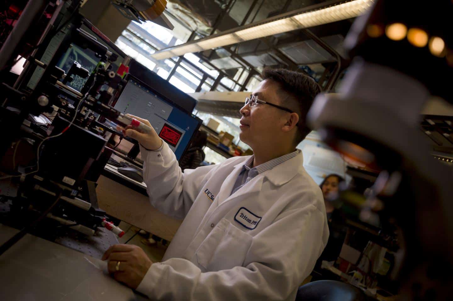
"We can directly print detailed microvasculature structures in extremely high resolution. Other 3D printing technologies produce the equivalent of 'pixelated' structures in comparison and usually require sacrificial materials and additional steps to create the vessels," said postdoc researcher Wei Zhu, a lead researcher on the project and co-lead author of a paper explaining the work in the journal Biomaterials.
Testing the technique, the team created a network containing endothelial cells, which line living blood vessels. They then implanted the structures, which measured 4 x 5 mm and were 600µm thick, into skin wounds on mice, finding that after two weeks they had merged with the mices’ circulatory systems and were supporting normal blood flow.
The vessels cannot yet transport nutrients and waste, Prof Chen notes. "We still have a lot of work to do to improve these materials,” he said. “This is a promising step toward the future of tissue regeneration and repair.”
The team is now working towards using patient-specific pluripotent stem cells to print blood vessel networks, but Chen warns that clinical trials are still some years off.


April 1886: the Brunkebergs tunnel
First ever example of a ground source heat pump?