
Scientists at Houston’s Rice University and The Methodist Hospital Research Institute (TMHRI) led an international team of researchers in creating composite particles that can be injected into patients and guided by magnetic fields. Once in position, the particles may be heated to kill malignant tissues or trigger the release of drugs at the site.
The so-called nanoconstructs should fully degrade and leave the body within a few days, they reported. The research appears online in the journal Advanced Functional Materials.
The team led by Rice chemist Lon Wilson and TMHRI scientist Paolo Decuzzi was searching for a way to overcome the challenges presented by iron oxide particles, which can be manipulated with magnets, provide excellent contrast under MRI, create heat when triggered and degrade quickly. They can’t, however, perform these tasks simultaneously so the team devised a way of decoupling the functions from their sizes.
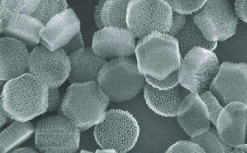
The solution was to package thousands of iron oxide particles – with magnetic cores as small as five nanometres across – inside larger particles.
The researchers made two such nanoconstructs, embedding iron oxide particles in silicon mesoporous particles (SiMPs) and discoidal polymeric nanoconstructs (DPNs). They knew from previous research that submicron-sized SiMPs and DPNs naturally accumulate within the tumour’s blood vessels.
Iron oxide enhances the ability to position and hold the particles in place with magnets, said lead author and Rice graduate student Ayrat Gizzatov.
‘They get attracted by the magnet, and that induces another dipole-dipole magnetic interaction among the particles and increases their interparticle communication mechanism,’ he said in a statement.
Tests showed iron oxide particles made the nanoconstructs 10 times better than traditional contrast agents with what amounted to significantly lower doses of iron than used in current practice.
The new research also showed that, as a general principle, confining MRI contrast agents (like iron oxide) in geometric structures enhances their relaxivity – the property that makes the agents appear in MRI images.
While the particles are too big to target specific proteins, Gizzatov said it might also be possible to modify them with elements that will increase their accumulation in tumours.


Red Bull makes hydrogen fuel cell play with AVL
Formula 1 is an anachronistic anomaly where its only cutting edge is in engine development. The rules prohibit any real innovation and there would be...