While still in the early stages, the research reported in Science Advances has taken steps towards a new MRI method with the potential to enable doctors to personalise medical treatments and allow real-time imaging to take place in operating theatres and GP practices.
MRI detects the magnetism of molecules to create an image and has become a crucial tool in medical diagnostics. However, a typical hospital scanner will effectively detect one molecule in every 200,000, making it difficult to see the full picture of what’s happening in the body.
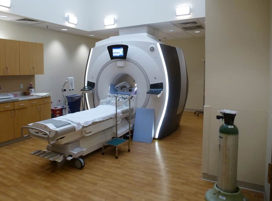
Improved scanners are now on trial in various countries, but because they operate in the same way as regular MRI scanners — using a superconducting magnet — these new models remain bulky and expensive.
The research team at York University has discovered a way to make molecules more magnetic and more visible, which could lead to a new generation of low-cost and highly sensitive imaging techniques.
The research team is said to have found a way to transfer the “invisible” magnetism of parahydrogen — a magnetic form of hydrogen gas—– into an array of molecules that occur naturally in the body such as glucose, urea and pyruvate. Using ammonia as a carrier, the researchers have been able to “hyperpolarise” substances such as glucose without changing their chemical composition, which would risk them becoming toxic.
According to York University, it is now theoretically possible that these magnetised, non-harmful substances could be injected into the body and visualised. Because the molecules have been hyperpolarized there would be no need to use a superconducting magnet to detect them as smaller, cheaper magnets would be able to do the same job.
If the method were to be successfully developed it could enable a molecular response to be seen in real time and the low-cost, non-toxic nature of the technique would introduce the possibility of regular and repeated scans for patients. These factors would improve the ability of the medical profession to monitor and personalise treatments, possibly resulting in more successful outcomes for individuals.
“In theory, it would provide an imaging technique that could be used in an operating theatre,” said Prof Simon Duckett from the Centre for Hyperpolarisation in Magnetic Resonance at York University. “For example, when a surgeon extracts a brain tumour from a patient they aim to remove all the cancerous tissue while at the same time removing as little healthy tissue as possible. This technique could allow them to accurately visualise cancerous tissue at a far greater depth there and then.”
The research also has the potential to bring MRI to countries in the developing world that don’t have the uninterrupted power supplies or infrastructure to operate current scanners. The method could also provide benefits to the chemical and pharmaceutical industries in addition to environmental and molecular science.


Red Bull makes hydrogen fuel cell play with AVL
Formula 1 is an anachronistic anomaly where its only cutting edge is in engine development. The rules prohibit any real innovation and there would be...