
‘Let’s say you have a large population of cells,’ said Corey Neu, an assistant professor in Purdue University’s Weldon School of Biomedical Engineering. ‘Just one of them might metastasize or proliferate, forming a cancerous tumour. We need to understand what it is that gives rise to that one bad cell.’
Such an advance makes it possible to simultaneously study the mechanical and biochemical behaviour of cells, said biomedical engineering postdoctoral fellow Charilaos ‘Harris’ Mousoulis.
Being able to study a cell’s internal workings in fine detail would likely yield insights into the physical and biochemical responses to its environment. The technology, which combines an atomic force microscope and nuclear magnetic resonance system, could help researchers study individual cancer cells, for example, to uncover mechanisms leading up to cancer metastasis for research and diagnostics.
The prototype’s capabilities were demonstrated by taking nuclear magnetic resonance spectra of hydrogen atoms in water. Findings represent a proof of concept of the technology and are detailed in a research paper that appeared in the journal Applied Physics Letters. The paper was co-authored by Mousoulis; research scientist Teimour Maleki; Babak Ziaie, a professor of electrical and computer engineering; and Neu.
‘You could detect many different types of chemical elements, but in this case hydrogen is nice to detect because it’s abundant,’ Neu said in a statement. ‘You could detect carbon, nitrogen and other elements to get more detailed information about specific biochemistry inside a cell.’
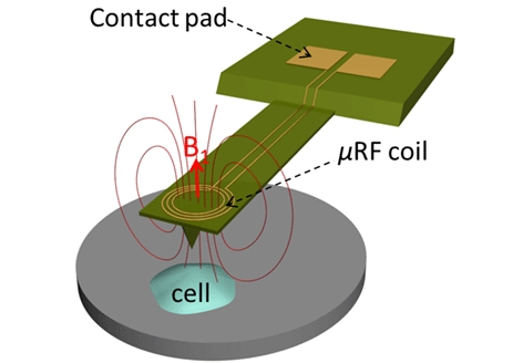
An atomic force microscope (AFM) uses a cantilever to yield information about materials and surfaces on the nanometre scale. Because the instrument enables scientists to observe objects far smaller than possible using light microscopes, it could be ideal for studying molecules, cell membranes and other biological structures.
However, the AFM does not provide information about the biological and chemical properties of cells so the researchers fabricated a metal microcoil on the AFM cantilever.
An electrical current is passed though the coil, causing it to exchange electromagnetic radiation with protons in molecules within the cell and inducing another current in the coil, which is detected.
A US patent application has been filed for the concept. The research has been funded by Purdue’s Showalter Trust Fund and the National Institutes of Health.


April 1886: the Brunkebergs tunnel
First ever example of a ground source heat pump?