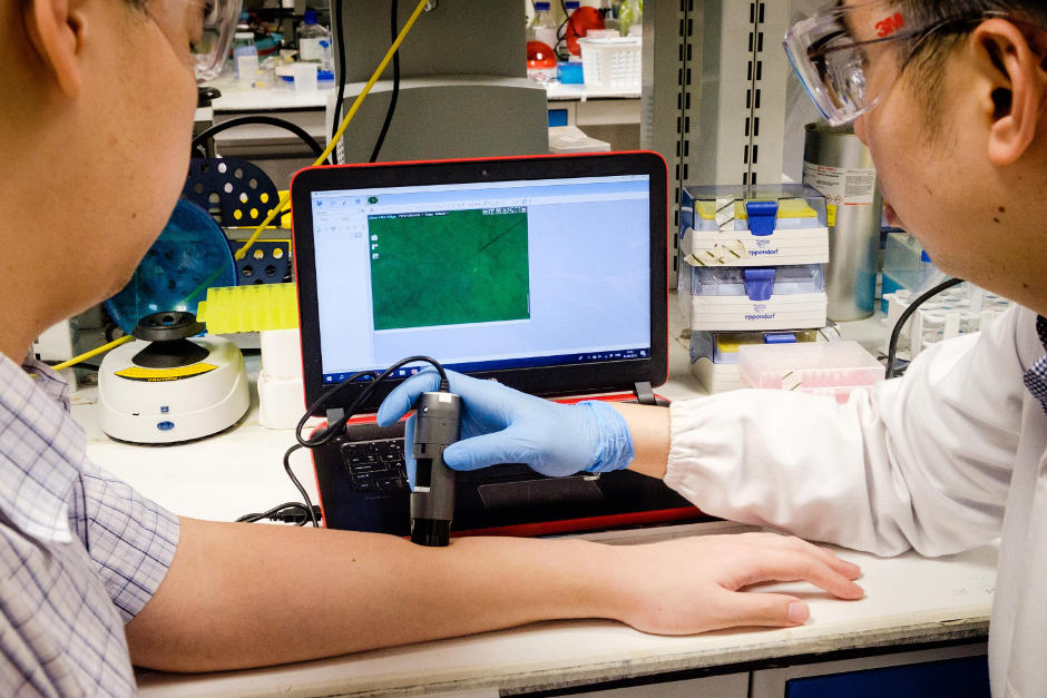
Dr David Yeo (right) using a microscope to detect the nanoflares
Clinicians currently find it difficult to predict how scars will develop without resorting to invasive testing.
Using the new nanoparticles, the joint research team from Nanyang Technological University, Singapore (NTU Singapore) and Northwestern University has shown in animals and human skin samples the potential to quickly and accurately predict whether a wound is likely to lead to excessive scarring.
If necessary, doctors can then take conventional preventive measures to reduce scar formation, such as using silicon sheets to keep a wound flat and moist.
The new technique was developed by a team led by Assistant Professor Xu Chenjie from NTU’s School of Chemical and Biomedical Engineering, nanoscience expert Professor Chad A Mirkin from Northwestern University, and Dr Amy S Paller, Chair of Dermatology at Northwestern University Feinberg School of Medicine.
Published in Nature Biomedical Engineering, the new detection method is said to use thousands of nanoparticles called NanoFlares, which have DNA strands attached to their surfaces like a ball of spikes.
These nanoparticles are applied to closed wounds using a cream. After the nanoparticles have penetrated the skin cells for 24 hours, a handheld fluorescence microscope is used to look for signals given out by the nanoparticles’ interaction with target biomarkers inside the skin cells.
If fluorescence signals are detected, they indicate abnormal scarring activity and preventive action can be taken to hopefully avoid heavier scarring.
Currently, apart from the visual examination of mature scars, the only other tool to detect skin diseases accurately is to perform a biopsy.
Assistant Professor Xu Chenjie said: “When our bioengineered nanoparticles are applied on the skin, they will penetrate up to 2mm below the skin surface and enter scar cells.”
“Upon binding with a specific tell-tale gene released by the scar cells, smaller DNA spikes are knocked loose and light up under the microscope like little light flares. The more flares we see, the more scarring activity there is.”
These NanoFlares are made by coating Northwestern’s patented gold nanoparticles with tiny DNA strands targeting particular genes. It has shown negligible toxicity or side effects when tested on mice, rabbits and on human skin samples.
Dr Amy S Paller, who is also the Director of Northwestern University Skin Disease Research Centre, said: “Beyond clinical observation, the gold standard for both clinical diagnosis and translational research of skin disorders is a biopsy.
“This technology is an exciting first step towards having a non-invasive way to detect increases or decreases in gene expression.”


April 1886: the Brunkebergs tunnel
First ever example of a ground source heat pump?