It will allow anaesthetists a moment-by-moment account of patients undergoing major surgery or for close monitoring of trauma, such as head injury or stroke.
Temporal brain scanning, which can see how the brain reacts to various stimuli, already exists in the form of functional magnetic resonance imaging (fMRI). This technology works by tracking changes in blood flow that are indicative of underlying neural activity. Yet because it based on the principle that active neurons demand oxygen, there is a natural lag in the time it takes blood flow to respond.
By contrast, the new imaging method — called functional electrical impedance tomography of evoked response (fEITER) — is based on rapid electrical activity deep in the brain.
The machine itself is a portable, lightweight monitor that can fit on a small trolley. It has 32 electrodes that are fitted around the patient’s head. A small, high-frequency electric current, too small to be felt or have any effect, is passed between two of the electrodes and the voltages between other pairs of electrodes are measured, in a process that takes less than one-thousandth of a second.
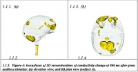
An electronic scan is thus carried out and the machine does this whole imaging procedure 100 times a second. By measuring the resistance to current flow, a 3D image of the changing electrical conductivity within the brain is constructed.
‘The electronics have been very challenging — to achieve this combination of sensitivity, low noise and high framing rate we’re using a very high-spec, in-house-designed current-injection system,’ said project lead Prof Hugh McCann, of the School of Electrical and Electronic Engineering at Manchester University.
The development and testing of fEITER was done in concert with a team of anaesthetists, also at Manchester, led by Prof Brian Pollard.
‘Anaesthetists are essentially consciousness engineers,’ said McCann. ‘It’s quite remarkable how deeply they understand the practical operation of the brain in terms of its degree of responsiveness… and it’s important they have good tools to monitor the degree of sedation.
‘Say a patient is admitted for a serious head injury, there’s a need to control things such as swelling of the brain — in that situation, anaesthetists are literally desperate to have more tools to monitor the situation and control it better.’
The fEITER project received funding from the Wellcome Trust in 2008, culminating in a recent clinical trial of 20 healthy volunteers and 20 anaesthetised patients scheduled for surgery.


Swiss geoengineering start-up targets methane removal
No mention whatsoever about the effect of increased methane levels/iron chloride in the ocean on the pH and chemical properties of the ocean - are we...