
While techniques such as computed tomography and magnetic resonance imaging have revolutionised medicine, they are not perfect – and it can be difficult to dig out the information that’s required.
’The paradigm that’s developed over time is simply to produce the best quality images possible, which are then interpreted by a specialist,’ said Hawkes. ’We believe that’s missing a trick. In many cases, we know the information we want, but producing a high-quality image and then assessing it is an indirect way of answering that question. We want to make extracting that clinically relevant information the driving force behind the whole imaging process.’
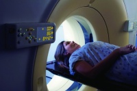
A scan takes several minutes and the patient has to lie still as the results are affected by movement. But when the scan is between the neck and the pelvis, there are many involuntary movements that cannot be suspended – from breathing and heartbeats to the digestive motion of the gut and the filling of the bladder. This motion corrupts the images, which tends to hide crucial information. For example, in a cancer patient, it can make it difficult to see the local blood supply to a tumour, or how densely packed its cells are.
The idea behind the intelligent imaging project is to optimise image acquisition to answer a specific question, rather than focusing solely on producing the best possible quality image. ’The information embedded in the data that’s used to reconstruct the image can be obscured or even thrown away in the reconstruction process, and it certainly gets obscured by patient motion,’ said Hawkes.
’Our plan is to take a computational model of the microstructure of what we’re looking for, such as a computer representation of the growth of the tissue of the abnormal blood supply or the abnormal division of cells, and then couple that with an understanding of how things are going to be moving around from respiration, cardiac motion and so on. Could this model then be coupled with the image acquisition to extract the precise information we want?’
The potential applications are widespread. Initially, the project will focus on three areas – cancer, cardiac ischaemia and studying what is happening to the brains of near-term foetuses, premature babies and those who have had difficult births. In all of these cases it is impossible to arrest motion to give a clear image, and this predictive model may be able to give much more detailed information about cancer cells, heart tissue or brain activity.
It also has potential in treatment, for example, in high-intensity focused ultrasound. Diagnostic ultrasounds have a negligible effect on tissue, but if the power is increased, sufficient energy is delivered to ’cook’ it. If this is then focused down to a point, it can be used to kill cells. Intelligent imaging could offer real-time information of how the treatment is progressing.
EPSRC has awarded the project team £6m over the next five years. Collaborators include magnetic resonance specialist Joe Hajnal at Imperial’s Hammersmith Hospital, cardiologist Reza Razavi at King’s College London, physicists Martin Leach and Steve Webb at the Institute for Cancer Research in Sutton, and Nic Smith from the computation lab at Oxford University.
The project timescale varies for different applications. Hawkes said it may be tested in prostate cancer within a year, whereas it will be two or three years for the liver. There are more problems to be solved in the cardiac and neonatal areas, so it may be three or four years before these reach the clinic.


Nanogenerator consumes CO2 to generate electricity
Whoopee, they've solved how to keep a light on but not a lot else.