Breast cancer diagnosis can take a number of weeks, involving a visit to the GP, followed by an x-ray mammogram and an ultrasound or even an MRI, before the patient finally undergoes a biopsy.
The new system, being developed by researchers at the University of Twente in the Netherlands, uses light and sound to detect signs of a tumour. The photoacoustic technique combines lasers and photonics with ultrasound.
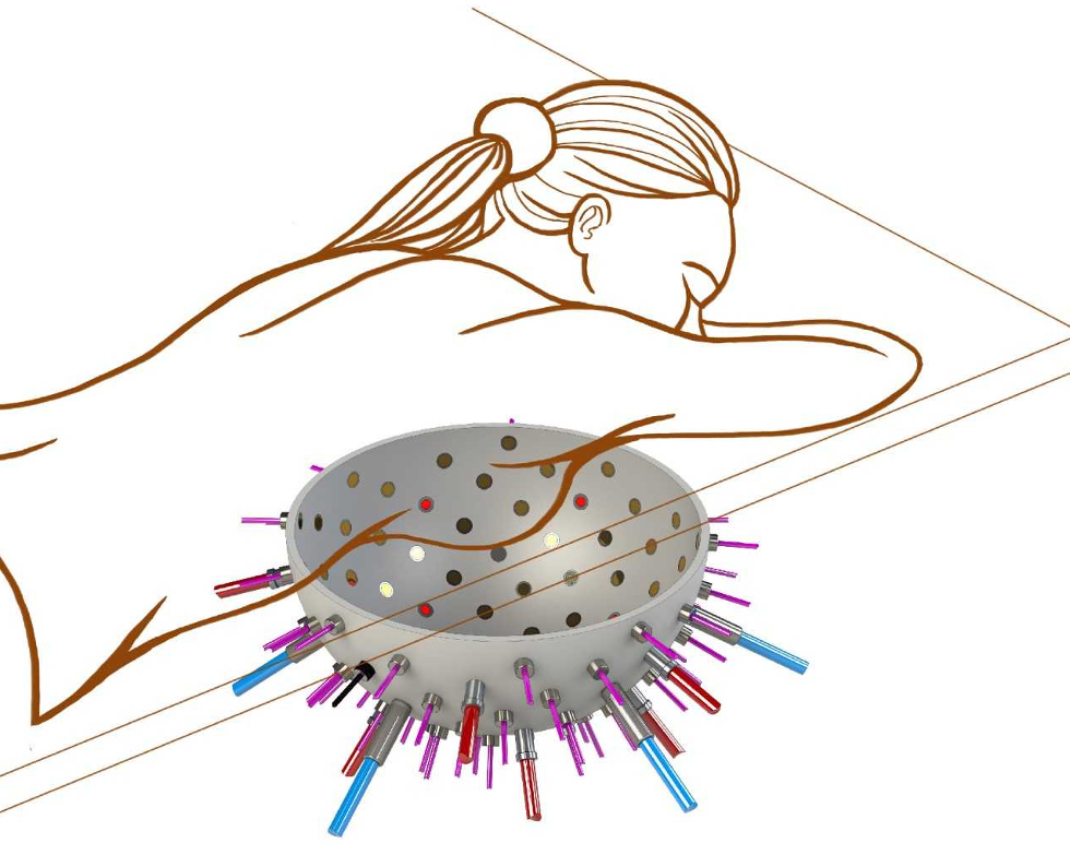
The patient lies face down on a bed, placing their breast into a hemispherical bowl lined with up to 100 optical fibres, and several ultrasound detectors.
Multiple images are taken from different angles. These are then assembled into a single 3D image, according to Srirang Manohar, the project coordinator.
“The imager will be non-invasive, will not require contrast agents nor use ionising radiation,” he said. “Furthermore, the patient will feel no pain or discomfort.”
The PAMMOTH (Photoacoustic Ultrasound Mammoscopy for evaluating screening-detected abnormalities in the breast) system operates by sending short pulses of light towards the suspect lesion.
“Light scatters within the breast and is selectively absorbed by blood in the strongly vascularised tumour site,” said Manohar. “This absorbed energy is converted into thermal energy and via thermal expansion into a pressure wave.”
This pressure wave can be picked up by the ultrasound detectors, he said. “From the detected signals the locations where the initial acoustic pressure was created can be reconstructed, which then gives a 3D map of the presence of tumour vasculature inside the breast.”
The system also analyses oxygen levels in the blood around the suspected tumour. Tumours consume oxygen at high rates in order to survive, so lower oxygenation levels around the lesion could indicate that it is malignant.
The system shines multi-wavelength light in the near-infrared range at the tissue. Oxygen coupled to haemoglobin in the blood absorbs light differently at certain wavelengths compared to blood with reduced haemoglobin.
Researchers at UCL in London are developing the mathematics, image reconstruction and signal analysis techniques to interpret the information and calculate how aggressive the tumour could be.
The on-going project has received a €4.35m grant from Horizon 2020. The PAMMOTH team hopes to have a prototype ready for 2020 ready for completion in 2021.


JLR teams with Allye Energy on portable battery storage
This illustrates the lengths required to operate electric vehicles in some circumstances. It is just as well few electric Range Rovers will go off...