
The Warwick researchers have been able to decorate the hollow structures of vesicles — microscopic polymer-based sacs of liquid — with a variety of nanoparticles that can give them ’stealth’ capabilities, which can avoid the body’s defences while releasing a drug.
Advances in polymerisation have led to a surge in the creation of vesicles made from polymer molecules. Such vesicles have interesting chemical and physical properties that make the hollow structures potential drug-delivery vehicles.
But the University of Warwick team, led by associate professor Stefan Bon, was convinced that even more strength, and interesting tailored properties, could be given to the vesicles if they could add an additional layer of colloidal armour made from a variety of nanoparticles.
One of those armour types was a highly regular packed layer of microscopic polystyrene balls. As a result of the crystalline-like ordered structure of the balls, the researchers could design a vesicle that had an additional and precise permeable reinforced barrier for drug release.
The researchers also succeeded in adding a gelatine-like polymer to provide a ’stealth’ armour to shield vesicles from unwanted attention from the body’s immune system while they slowly released drugs. That particular coating absorbs so much surrounding water into its outer structure that it may be able to fool the body’s defence mechanism into believing it is just water.
The Warwick researchers also had the idea of simply giving their chosen colloidal particles the opposite charge to that of the polymer vesicles.
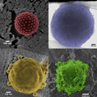
However, the researchers needed a new way of actually observing the vesicles to see if their plan had worked. Previous observational methods required researchers to dry out the vesicles before examining them under an electron microscope — but this seriously deformed the vesicles and therefore provided little useful data.
However, by using a cryo-electron microscope, the research team could quickly freeze the vesicles to -150oC, preserving their shape, before observation by the electron microscope to show that the simple charge-based method had worked as planned.


Nanogenerator consumes CO2 to generate electricity
Whoopee, they've solved how to keep a light on but not a lot else.