Researchers have used sound waves to separate circulating tumour cells from blood samples quickly and efficiently, a development that could lead to more accurate liquid biopsies.
CTCs are small pieces of a tumour that break away and flow through the bloodstream. They contain information such as tumour type, physical characteristics and genetic mutations.
The ability to quickly and efficiently harvest and grow these cells from a blood sample would enable liquid biopsies capable of providing robust diagnosis, prognosis and suggestions for treatment strategies based on individual CTC profiling.
Catching CTCs is difficult as there are typically only a handful for every few billion blood cells running through a patient's veins. Technologies that separate tumour cells from normal blood cells already exist but they tend to damage or kill the cells in the process, lack efficiency, only work on specific types of cancer, or take too long to be used in many situations.
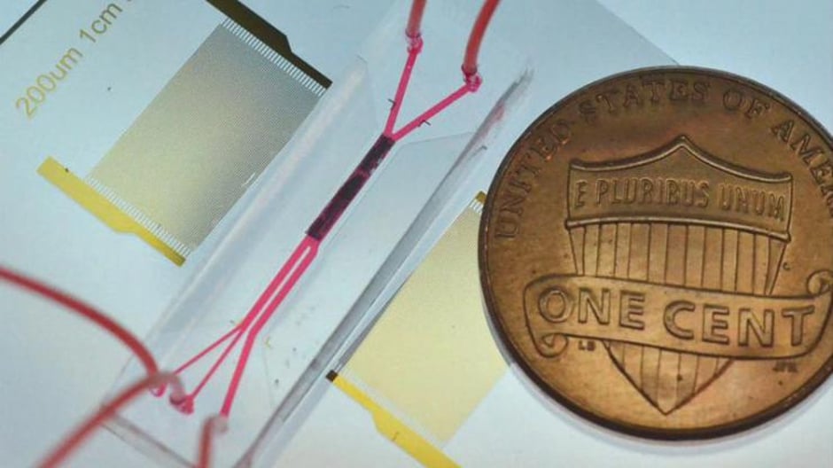
In a new study, researchers from Duke University, MIT and Nanyang Technological University in Singapore demonstrate a platform based on sound waves that is reportedly capable of separating CTCs from a 7.5mL vial of blood with at least 86 per cent efficiency in under an hour. With additional improvements, the researchers hope the technology will form the basis of a new test through an inexpensive, disposable chip.
The results have been published in the journal Small.
"Biopsy is the gold standard technique for cancer diagnosis," said Tony Jun Huang, the William Bevan Professor of Mechanical Engineering and Materials Science at Duke. "But it is painful and invasive and is often not administered until late in the cancer's development. With our circulating tumour cell separation technology, we could potentially help find out, in a non-invasive manner, whether the patient has cancer, where the cancer is located, what stage it's in, and what drugs would work best. All from a small sample of blood drawn from the patient."
https://www.youtube.com/watch?v=XdmCK8AflZk
The technology works by setting up a standing sound wave at an angle to a fluid flowing through a tiny channel. This sets up pockets of pressure that push on particles suspended in the liquid as they pass. This acoustic force acts more strongly on the larger, more rigid cancer cells than on normal blood cells, pushing the CTCs into a separate channel for collection.
The power intensity and frequency of the sound waves are similar to those used in ultrasonic imaging. The risk of damage to the CTCs is reduced even further because each cell experiences the acoustic wave for a fraction of a second and does not require labelling or surface modification. These features are said to give the technique the best possible chance at maintaining the functions and native states of the CTCs.
The approach was first demonstrated three years ago in a proof-of-concept study and has since been improved to the point where it could be useful in a clinical setting. The result is a prototype device that processes fluid at a rate of 7.5mL/hour, seven times faster than the original, without sacrificing any of its 86 per cent efficiency or numerous advantages over other methods.
"The biggest asset of this acoustic method of separation is that it's very gentle on the circulating tumour cells," said Andrew Armstrong, associate professor of medicine, surgery, and pharmacology and cancer biology at the Duke University School of Medicine. "The cancer cells remain viable after passing through the chip and can be characterised, cultured or profiled, which allows us to do genotyping or phenotyping to better understand how to kill them."


Project to investigate hybrid approach to titanium manufacturing
What is this a hybrid of? Superplastic forming tends to be performed slowly as otherwise the behaviour is the hot creep that typifies hot...