
The breakthrough, made by researchers at Shanghai University’s Rapid Manufacturing Engineering Center, has allowed researchers to utilise separate materials that possess mechanical strength and promote new cell growth, a problem for existing vascular grafts that have consisted of a single or double layer.
Vascular grafts are surgically attached to an obstructed or otherwise unhealthy blood vessel to permanently redirect blood flow, such as in coronary bypass surgery. Traditional grafts work by repurposing existing vessels from the patient’s body or from a suitable donor.
However, these sources are often insufficient for a patient’s needs because of the limited supply in a patient’s body, and may be afflicted by the same underlying conditions that necessitate the graft in the first place.
Accordingly, there has been a great deal of research towards developing synthetic vessels that can mimic natural ones, allowing new cells to grow around them and then degrade away, thereby creating new vessels.
‘The composite vascular grafts could be better candidates for blood vessel repair,’ said Yuanyuan Liu, an associate professor at the Rapid Manufacturing Engineering Center. Liu and her team describe their current research in the AIP Advances.
According to the American Institute of Physics, surrogate scaffolds usually need to mimic the natural vasculature of their targeted tissue as much as possible. For blood vessel surrogates, this structural mimicry can be fabricated by electrospinning, a process which uses an electrical charge to draw liquid inputs, in this case a mixture of chitosan and polyvinyl alcohol – into incredibly fine fibres.
Electrospinning also allows for a high surface-to-volume ratio of Nano fibres, providing ample space for host cells to grow and connect. These components all naturally degrade within six months to a year, leaving behind a new, intact blood vessel.
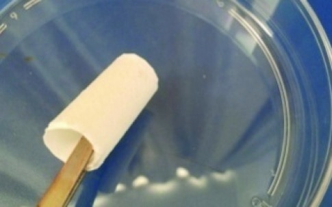
The resulting structure, however, isn’t very rigid so to compensate for this, the researchers are said to have designed a three-layer model, in which the mixture was electrospun onto both sides of a microimprinted middle layer of poly-p-dioxanone, a biodegradable polymer commonly used in biomedical applications. The ends of this sheet were then folded and attached to make a tube-like vessel.
Liu and her team then seeded the scaffold with rat fibroblast cells to test the scaffold’s efficacy in promoting cellular expansion and integration. The researchers found that the cells on these composite scaffolds proliferated quickly, likely due to the functional amino and hydroxyl groups introduced by the chitosan.
While work remains before the prospect of human trials, Liu and her group are optimistic about the future of their research. Their next project is to test the implants in an animal model, to observe the structure’s efficacy with live vascular cells.


Nanogenerator consumes CO2 to generate electricity
Whoopee, they've solved how to keep a light on but not a lot else.