The technique, known as phase-contrast X-ray imaging, has been developed in a five-year, EPSRC-funded project, led by University College London (UCL).
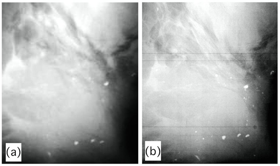
Rather than measuring the extent to which tissue or materials absorb radiation - as in conventional X-ray imaging – the technique measures the physical effect that passing through different types of tissue or material has on the speed of the X-ray itself.
The technique can also spot smaller cracks and defects in materials than conventional X-rays, as it is excellent at determining different shapes and types of matter – something conventional X-rays could only match by using prohibitively high doses of radiation.
The technique has previously been limited to large-scale synchrotron facilities such as Oxfordshire's Diamond Light Source, according to UCL’s Professor Alessandro Olivo, who led the project.
“We've now advanced this embryonic technology to make it viable for day-to-day use in medicine, security applications, industrial production lines, materials science, non-destructive testing, the archaeology and heritage sector, and a whole range of other fields,” he said.
The technology has been incorporated into a prototype security scanner by Nikon Metrology, designed to detect weapons and explosives concealed in baggage, for example.
And in a new three-year project, supported by the Wellcome Trust, the Nikon Metrology/UCL team will develop a prototype scanner for breast cancer surgery in collaboration with Barts Health and Queen Mary University of London, which should help surgeons determine the exact extent of the malignancy and reduce the need to recall patients for further operations.
The technology can also detect some tissue types invisible to conventional X-ray machines, such as cartilage, and there are plans to set up a spinout company to commercialise this aspect of the technology.


Nanogenerator consumes CO2 to generate electricity
Whoopee, they've solved how to keep a light on but not a lot else.