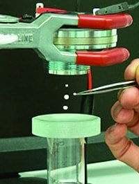
Using ultrasound, cartilage cells taken from a patient’s knee can be levitated for weeks in a nutrient-rich fluid. In doing so, the nutrients can reach every part of the culture’s surface and, combined with the stimulation provided by the ultrasound, enables the cells to grow and to form better implant tissue than when grown in a petri dish.
By holding the cells in the required position, the tweezers can also mould the growing tissue into the right shape so that the implant is fit-for-purpose when inserted into the patient’s knee. Over 75,000 knee replacements are carried out each year in the UK and many could be avoided if cartilage implants could be improved.
The ultrasonic tweezers were developed by researchers from the Universities of Southampton, Bristol, Dundee and Glasgow along with industrial partners.
Prof Martyn Hill, head of the Engineering Sciences Unit at the University of Southampton, led the cartilage tissue engineering work in collaboration with colleagues Dr Peter Glynne-Jones, New Frontiers Fellow in Engineering Sciences, Dr Rahul Tare, a Lecturer in Musculoskeletal Science and Bioengineering, and Professor Richard Oreffo, a Professor of Musculoskeletal Science.
In a statement, Prof Hill said: ‘Ultrasonic tweezers can provide what is, in effect, a zero-gravity environment perfect for optimising cell growth. As well as levitating cells, the tweezers can make sure that the cell agglomerates maintain a flat shape ideal for nutrient absorption. They can even gently massage the agglomerates in a way that encourages cartilage tissue formation.’
Prof Bruce Drinkwater of Bristol University, who co-ordinated the programme, said: ‘Ultrasonic tweezers have all kinds of possible uses in bioscience, nanotechnology and more widely across industry. They offer big advantages over optical tweezers relying on light waves and also over electromagnetic methods of cell manipulation; for example, they have a complete absence of moving parts and can manipulate not just one or two cells at a time but clusters up to 1mm across – a scale that makes them very suitable for applications like tissue engineering.’
The tweezers, developed with Engineering and Physical Sciences Research Council (EPSRC) funding, involve multiple, tiny beams of ultrasonic waves that, in a typical device, point into a 10mm-diameter chamber. With the aid of a microscope to monitor the procedure, the forces generated by the waves can then be manipulated so that they move cells into the required position, turn them around, or hold them firmly in place.
The research programme has also shown that ultrasonic tweezers can be used to build up cell tissue layer by layer, which could reconstruct nerve tissue after severe trauma such as limb amputation.
According to Southampton University, this research will enable ultrasonic tweezer technology to be refined and miniaturised and specific uses to be explored and developed in the next few years. The first real-world applications, in sectors including bioscience and electronics, could potentially be developed within around five years.


Swiss geoengineering start-up targets methane removal
No mention whatsoever about the effect of increased methane levels/iron chloride in the ocean on the pH and chemical properties of the ocean - are we...