
It is claimed that the X-ray system, developed by Prof Robert Cernik and colleagues from the university’s School of Materials, can identify chemicals and compounds such as cocaine, semtex, precious metals or radioactive materials, even when concealed in large objects.
The method could also be extended to detect strain in fabricated components such as aircraft wings and it can be used to image corrosion processes and chemical changes.
In healthcare, the system can be used to detect abnormal tissue types from biopsy samples, while it could be employed in geophysical exploration to analyse the content of core samples taken from bore holes.
In a recent experiment, the team used the technology to X-ray a USB dongle that controls webcams. The team was able to identify the different elements and components inside the dongle by analysing the energy-sensitive radiographs and fluorescence patterns.
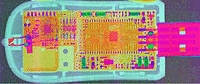
The elements or components — bromine, barium, silver, tin and zirconium — were highlighted in different colours to identify them to the system operators. The results of the tests have been published in the journal Analyst.
In a statement, Prof Robert Cernik said: ‘The fact that we can now use this technology in a laboratory setting is a substantial step forward.
‘When we first developed the idea five years ago, we needed the power of a synchrotron to produce the X-rays. In addition, we only had access to silicon-based detectors.
‘This is a problem because silicon is a light atom and will not stop the high-energy X-rays that come through large objects. Now we can achieve the same imaging results with an 80 x 80-pixel camera [made from cadmium zinc telluride] that supports real-time hyperspectral X-ray imaging up to very high energies.
‘Current imaging systems such as spiral CAT scanners do not use all the information contained in the X-ray beam. We can use all the wavelengths present to give a colour X-ray image in a number of different imaging geometries.
‘This method is often called hyperspectral imaging because it gives extra information about the material structure at each voxel of the 3D image. This extra information can be used to fingerprint the material present at each point in a 3D image.’
As well as providing more information about the object being X-rayed, this new technique is claimed to decrease the time it takes to create a 3D image.
Rather than building up lots of separate images, the new system creates the image in one scanning motion that now only takes several minutes.
Cernik is now seeking industrial partners for collaborative projects to refine the X-ray technology for each specific application, including security, aerospace and medical imaging.


Nanogenerator consumes CO2 to generate electricity
Nice to see my my views being backed up by no less a figure than Sabine Hossenfelder https://youtu.be/QoJzs4fA4fo