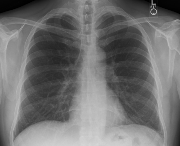
Chest X-rays account for 40 per cent of all diagnostic imaging worldwide, and it’s estimated that at any given time in the UK there are over 300,000 X-rays that have been waiting more than 30 days for a report. The new system, built by researchers from Warwick University in collaboration with the NHS, is claimed to reduce the average time for a radiologist report on critical chest X-rays from 11 to three days.
“The increasing clinical demands on radiology departments worldwide has challenged current service delivery models, particularly in publicly-funded healthcare systems,” said research lead Professor Giovanni Montana, chair in Data Science at Warwick’s WMG (Warwick Manufacturing Group).
“It is no longer feasible for many radiology departments with their current staffing level to report all acquired plain radiographs in a timely manner, leading to large backlogs of unreported studies.”
The researchers extracted a dataset of half a million anonymised adult chest X-rays and developed an AI system for computer vision that can recognise radiological abnormalities in real-time, then suggest triage prioritisation by a radiologist. In the process of building the AI system, the team developed a Natural Language Processing (NLP) algorithm that can read a radiological report, understand the radiologist’s findings and automatically infer the priority level of the exam. They then applied this algorithm to the historical X-rays, training the AI to understand which visual patterns were predictive of their urgency level. It was found that normal chest radiographs were detected with a positive predicted value of 73 per cent and a negative predicted value of 99 per cent. The research is reported in the journal Radiology.

"The NLP goes well beyond pattern matching," said Professor Montana (right). "It uses AI techniques to infer the structure of each written sentence; for instance, it identifies the presence of clinical findings and body locations and their relationships. The development of the NLP system for labelling chest X-rays at scale was a critical milestone in our study.
“The results of this research shows that alternative models of care, such as computer vision algorithms, could be used to greatly reduce delays in the process of identifying and acting on abnormal X-rays - particularly for chest radiographs which account for 40 per cent of all diagnostic imaging performed worldwide. The application of these technologies also extends to many other imaging modalities including MRI and CT.”


First seven members join NG’s Great Grid Partnership
Agreed. It is all pretentious posturing and trite branding with no meaning or gravitas. Prepare to be disappointed by all of these greats/grates.