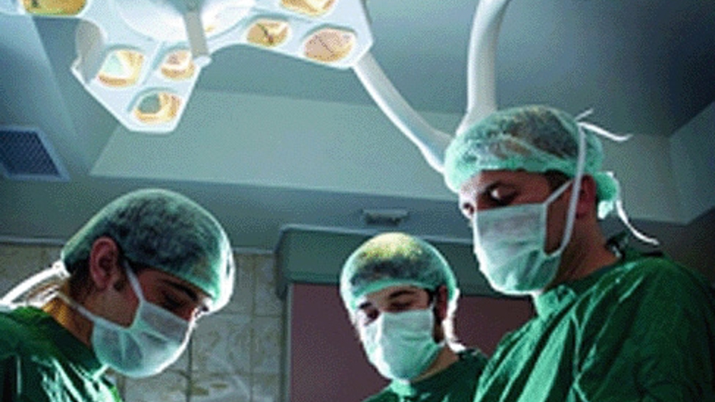
3D imagery helps surgeons plan surgery
Researchers at Jefferson Medical College in Philadelphia have developed a hologram-like display of a patient’s organs that surgeons can use to plan surgery.

This approach uses molecular PET/CT images of a patient to rapidly create a 3D image of that patient, so that surgeons can see the detailed anatomical structure, peel away layers of tissue, and move around in space to see all sides of a tumour, before entering the operating room to excise it.
‘Our technology presents PET/CT data in an intuitive manner to help physicians make critical decisions during surgical planning,’ said Matthew Wampole, Ph.D., from the Department of Biochemistry and Molecular Biology at Jefferson.
The researchers produced a surgical simulation of human pancreatic cancer reconstructed from a patient’s PET scans and contrast-enhanced CT scans. Six Jefferson surgeons evaluated the 3D model for accuracy, usefulness, and applicability of the model to actual surgical experience.
According to a statement, the surgeons reported that the 3D imaging technique would help in planning an operation. Furthermore, the surgeons indicated that the 3D image would be most useful if it were accessible in the operating room during surgery. The 3D image is designed to speed the excision of malignant tissue, avoiding bleeding from unusually placed arteries or veins, according to the report published September 24th in PLOS ONE.
Register now to continue reading
Thanks for visiting The Engineer. You’ve now reached your monthly limit of news stories. Register for free to unlock unlimited access to all of our news coverage, as well as premium content including opinion, in-depth features and special reports.
Benefits of registering
-
In-depth insights and coverage of key emerging trends
-
Unrestricted access to special reports throughout the year
-
Daily technology news delivered straight to your inbox


Water Sector Talent Exodus Could Cripple The Sector
Maybe if things are essential for the running of a country and we want to pay a fair price we should be running these utilities on a not for profit...