Down in the mouth

Researchers in the
Professor Keith Horner and Dr Hugh Devlin coordinated a three year, EU-funded collaboration with the Universities of Athens, Leuven,
Wide-scale screening for osteoporosis is not currently viable, largely due to the cost and scarcity of specialist equipment and staff.
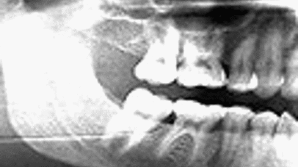
The team developed a software-based approach to detecting osteoporosis during routine dental X-rays, by automatically measuring the thickness of part of the patient’s lower jaw.
X-rays are used widely in the NHS to examine wisdom teeth, gum disease and during general check-ups, and their use is on the rise. In 2005 almost 6000 were taken on female patients aged 65 or over in a single month, and the number taken has increased by 181 per cent since 1981.
Register now to continue reading
Thanks for visiting The Engineer. You’ve now reached your monthly limit of news stories. Register for free to unlock unlimited access to all of our news coverage, as well as premium content including opinion, in-depth features and special reports.
Benefits of registering
-
In-depth insights and coverage of key emerging trends
-
Unrestricted access to special reports throughout the year
-
Daily technology news delivered straight to your inbox


British Steel Signs Five Year Deal With Network Rail
It was all very well to pass emergency legislation which amounted to a ´command economy´ for the industry (directors could be ordered to continue the...