
The method could be used to take precise three-dimensional images of plaques lining arteries, said Ji-Xin Cheng, an associate professor of biomedical engineering and chemistry at Purdue University.
Other imaging methods that provide molecular information are unable to penetrate tissue deep enough to reveal the three-dimensional structure of the plaques, but being able to do so would make better diagnoses possible, he added.
‘You would have to cut a cross-section of an artery to really see the three-dimensional structure of the plaque,’ Cheng said. ‘Obviously, that can’t be used for living patients.’
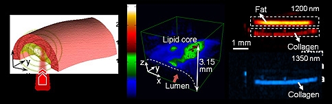
According to Purdue, the imaging reveals the presence of carbon-hydrogen bonds making up lipid molecules in arterial plaques that cause heart disease.
It is claimed that the method might also be used to detect fat molecules in muscles to diagnose diabetes and other lipid-related disorders, including neurological conditions and brain trauma. The technique also reveals nitrogen-hydrogen bonds making up proteins, meaning the imaging tool also might be useful for diagnosing other diseases and to study collagen’s role in scar formation.
‘Being able to key on specific chemical bonds is expected to open a completely new direction for the field,’ Cheng said
Findings are detailed in a paper to be published in Physical Review Letters. The findings represent the culmination of four years of research led by Cheng and doctoral student Han-Wei Wang.
The new technique is said to use nanosecond laser pulses in the near-infrared range of the spectrum.
The laser generates molecular ‘overtone’ vibrations, or wavelengths that are not absorbed by the blood. The pulsed laser causes tissue to heat and expand locally, generating pressure waves at the ultrasound frequency that can be picked up with a device called a transducer.
‘We are working to miniaturise the system so that we can build an endoscope to put into blood vessels using a catheter,’ Cheng said. ‘This would enable us to see the exact nature of plaque formation in the walls of arteries to better quantify and diagnose cardiovascular disease.’
The Purdue researchers are said to be the first to show that a strong photoacoustic signal can arise from the absorption of light by the chemical bonds in molecules. The near-infrared laser causes enough heating to generate ultrasound but not enough to damage tissue.
‘You can measure the time delay between the laser and the ultrasound waves, and this gives you a precise distance, which enables you to image layers of the tissues for three-dimensional pictures,’ Cheng added. ‘You do one scan and get all the cross-sections. Our initial target is cardiovascular disease, but there are other potential applications, including diabetes and neurological conditions.’
The approach is said to represent a major improvement over another imaging technique, coherent anti-Stokes Raman scattering (CARS), which has been used by the Purdue-based laboratory to study three-dimensional plaque formation in arteries.


Poll: Should the UK’s railways be renationalised?
I think that a network inclusive of the vehicles on it would make sense. However it remains to be seen if there is any plan for it to be for the...