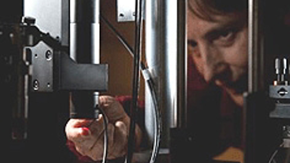
Liquid lens provides 3D images from under the skin's surface
University of Rochester optics professor Jannick Rolland has developed an optical technology that is claimed to provide unprecedented images from under the skin’s surface.

The aim of the technology is to detect and examine skin lesions to determine whether they are benign or cancerous without having to take a skin biopsy.
Instead, the tip of a cylindrical probe is placed in contact with the tissue and a clear, high-resolution 3D image of what lies below the surface quickly emerges.
‘My hope is that, in the future, this technology could remove significant inconvenience and expense from the process of skin-lesion diagnosis,’ said Rolland. ‘When a patient walks into a clinic with a suspicious mole, for instance, they wouldn’t necessarily have to have it surgically cut out of their skin or be forced to have a costly and time-consuming MRI done.
‘Instead, a relatively small, portable device could take an image that will assist in the classification of the lesion right in the doctor’s office.’
According to a statement from Rochester, the device accomplishes this using a liquid lens set-up developed by Rolland and her team for a process known as Optical Coherence Microscopy.
Register now to continue reading
Thanks for visiting The Engineer. You’ve now reached your monthly limit of news stories. Register for free to unlock unlimited access to all of our news coverage, as well as premium content including opinion, in-depth features and special reports.
Benefits of registering
-
In-depth insights and coverage of key emerging trends
-
Unrestricted access to special reports throughout the year
-
Daily technology news delivered straight to your inbox


Simulations show Optimal Design for Bladeless Wind Turbines
"an 80cm mast" Really? I'm short but that's only half my height! Do they mean 800cm?