Researchers investigating the optical properties of metamaterials claim to have created a lens which can potentially image single molecules and to detect cancerous cells. Made of minute slivers of gold and a transparent polymer, the lens works with visible light and is claimed to avoid the problems of refraction seen with conventional optical lenses.
Conventional optics reach the limit of their usefulness when the objects they are being used to study are of a similar scale to the wavelength of the light being used to illuminate them. Metamaterials — which have optical properties not found in nature — can avoid this problem by converting so-called ‘evanescent’ waves of light, which results from diffraction and is lost in conventional optics, into propagating light that can be collected and transmitted with standard equipment. Known as hyperlenses, early versions of these systems were composed of concentric rings of silver and an insulating material; the lens would be formed by slicing a complete circle of these rings along a diameter, with the light then entering along the curved side. However, these hyperlenses only worked with a narrow range of wavelengths and suffered large losses.
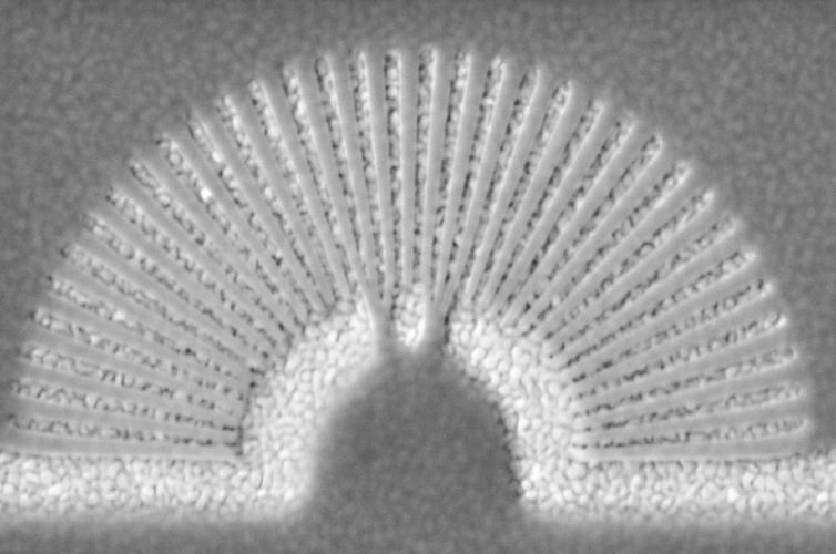
The new research, from at team led by Prof Natalia Litchinitser of the University of Buffalo, takes a different approach. Rather than using strips arranged concentrically, the team used alternating slivers of gold and the transparent acrylic polymer PMMA arranged radially around a semicircle. Silver or titanium nitride can be used in place of gold, while magnesium fluoride can be substituted for the PMMA. Each sliver is around 300nm thick.
In a paper in the journal Nature Communications, the team explains how the fan-shaped hyperlens can be made using electron beam lithography and electroplating, and integrated with a waveguide to perform optical experiments. The assembly worked with visible light and indicated that objects down to 250nm could be imaged, contrasting with around 10,000nm for the best optical endoscopes currently available.
“There is a great need in healthcare, nanotechnology and other areas to improve our ability to see tiny objects that elude even the most powerful optical systems. The hyperlens we are developing is, potentially, a giant step toward solving this problem,” Litchinitser said in a statement. For example, she notes in the paper, it could help visualise individual cancer cells in ovarian cancer biopsies, or adenocarcinoma, which can occur in the throats of people suffering from reflux disease: both are very hard to detect early with current diagnostics. Other applications include imaging single molecules in drug development, and manufacturing very small electronic components in semiconductors by using light to etch through a mask onto polymer film.


Poll: Should the UK’s railways be renationalised?
I think that a network inclusive of the vehicles on it would make sense. However it remains to be seen if there is any plan for it to be for the...