The team said the device - which uses so-called 'magic angle' effect - could potentially help diagnose knee injuries more quickly, and more accurately.
In a proof-of-concept study using animal knees, the results suggest the technology could be used to show all the structures of the knee.
The scientists say the device could help diagnose conditions such as anterior cruciate ligament injuries and its small size means it could eventually be used in local clinics and GP surgeries.
Dr Karyn Chappell, a researcher and radiographer from Imperial's MSK Lab said: "Knee injuries affect millions of people - and MRI scans are crucial to diagnosing the problem, leading to quick and effective treatment. However we currently face two problems: connective tissue in the knee is unclear on MRI scans, and people are waiting a long time for a scan.
"This can cause particular problems for women, as they are at greater risk of anterior cruciate ligament injuries. The reasons for this are unclear, but it could be linked to hormones such as oestrogen making ligaments more elastic, leading to more joint injuries."
Knee injuries commonly affect one of three areas: the tendons, the meniscus, or the ligaments. Following knee injury a doctor may refer a patient for a MRI scan to help establish which part of the joint is injured. MRI scans use a combination of radio waves and strong magnets to 'flip' water molecules in the body. The water molecules send out a signal, which creates an image.
However, tendons, ligaments and meniscus are not usually visible with MRI, due to the way water molecules are arranged in these structures, Dr Chappell said.
To overcome this problem, Dr Chappell harnessed the power of a phenomenon called the 'magic angle'. "The brightness of these tissues such as tendons and ligaments in MRI images strongly depends on the angle between the collagen fibres and the magnetic field of the scanner. If this angle is 55 degrees the image can be very bright, but for other angles it is usually very dark."
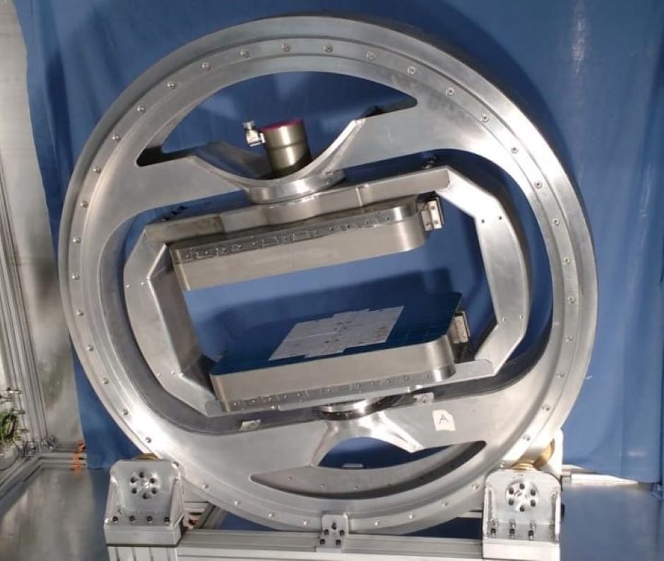
The team said the magic angle is achieved in their scanner because they are able to easily change the orientation of the magnetic field. While the patient sits in a chair, the specially designed magnet can rotate around the leg and the orientate magnetic field in multiple directions. This is not possible in current hospital MRI scanners, which are also much more expensive than the prototype scanner.
"Previously the magic angle phenomenon was thought of as a problem, as it could mean medical staff mistakenly thinking the knee is injured. However, I realised that if we took a number of scans around the knee, we could use the signal produced by the magic angle effect to build a clear picture of the knee structures," said Dr Chappell.
In a new study, published in Magnetic Resonance in Medicine, the multi-disciplinary team scanned the knee joints of six goats and ten dogs in a conventional MRI scanner.
Dogs suffer from knee injuries and arthritis similar to humans, making them a good subject for the study. The results showed that using the magic angle can accurately detect ligament and tendon damage.
The team say now they know magic angle scanning can be used to visualise the knee, combining this with the new prototype mini scanner could enable knees to be accurately scanned with this technology - and hope to progress to human trials of the 'mini' scanner within a year.
The research was funded by the National Institute for Health Research.


Poll: Should the UK’s railways be renationalised?
Rail passenger numbers declined from 1.27 million in 1946 to 735,000 in 1994 a fall of 42% over 49 years. In 2019 the last pre-Covid year the number...