The advance by a team at Nottingham University is the first of its kind in the world and could help to identify future drought- and flood-resistant crops.
The inability to monitor water uptake inside roots - without damaging the specimen - has been a key stumbling block for researchers seeking to understand the motion of fluids in living plant cells and tissues.
MORE FROM ENERGY & ENVIRONMENT
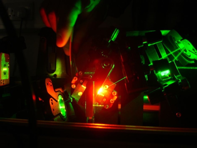
In a statement, study lead Dr Kevin Webb from the Optics and Photonics Research Group, said: “To observe water uptake in living plants without damaging them, we have applied a sensitive, laser-based, optical microscopy technique to see water movement inside living roots non-invasively, which has never been done before.
“Fundamentally, the process by which plants are able to thrive and become productive crops is based on how well it can take up water and how well it can manage that process. Water plays an essential role as a solvent for nutrients, minerals and other biomolecules in plant tissues. We’ve developed a way to allow ourselves to watch that process at the level of single cells. We can not only see the water going up inside the root, but also where and how it travels around.”
According to Nottingham University, water transport measurements were performed on the roots of Arabidopsis thaliana, which is a model plant for scientists because they can be genetically-engineered to interfere with basic processes like water uptake.
The new imaging technique - based on Raman scattering - allowed researchers to measure water travelling up through the root system of Arabidopsis at the cellular level, and to run a mathematical model to explain and quantify this.
The researchers used ‘heavy’ water (deuterium oxide), which contains an extra neutron in the nucleus of each hydrogen atom. By scanning a laser in a line across the root while the plant drank, it was possible to see the ‘heavy’ water moving past via the root tip.
In Arabidopsis that had been genetically-altered to compromise its water uptake, these measurements - combined with the mathematical model - revealed an important water barrier within the root. This confirmed for the first time that water uptake is restricted within the central tissues of the root, inside of which the water vessels are located.
Co-lead, Malcolm Bennett, Professor of Plant Sciences at the University, said, “This innovative technique is a real game-changer in plant science – enabling researchers to visualise water movement at a cell and second scale within living plant tissues for the very first time. This promises to help us address important questions such as – how do plants ‘sense’ water availability? Answers to this question are vital for designing future crops better adapted to the challenges we face with climate change and altered weather patterns.”
The findings of this Leverhulme Trust-funded study are published in Nature Communications.
The researchers are working towards a commercial path for their hydrodynamic Raman imaging technique, and have applied for funding with four UK and EU agriculture companies to look at tracers that move from plant leaves to roots to understand both directions of water transport. In parallel, the team is working on portable versions of the technology to allow water transport measurements to be taken into the field by farmers and scientists to monitor water handling in crops growing in challenging local environments.


Poll: Should the UK’s railways be renationalised?
The term innovation is bandied about in relation to rail almost as a mantra. Everything has to be innovative. There is precious little evidence of...