Rethinking evolution

The detailed images of embryos more than 500 million years old have been revealed by an international team of scientists, led by the
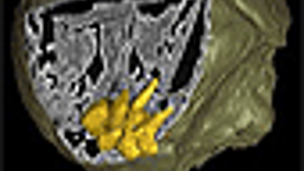
Writing in Nature, Dr Phil Donoghue and colleagues reveal the various developmental stages of fossilised embryos, from the first splitting of cells to pre-hatching, using synchrotron-radiation X-ray tomographic microscopy (SRXTM).
In one instance this has exposed the internal anatomy of the mouth and anus of a close relative of the living penis worm. Another case has revealed a unique pattern for making embryonic worm segments, not seen in any animals living today.
The
‘They are just gelatinous balls of cells that rot away within hours. But these fossils are the most precious of all because they contain information about the evolutionary changes that have occurred in embryos over the past 500 million years.’
Register now to continue reading
Thanks for visiting The Engineer. You’ve now reached your monthly limit of news stories. Register for free to unlock unlimited access to all of our news coverage, as well as premium content including opinion, in-depth features and special reports.
Benefits of registering
-
In-depth insights and coverage of key emerging trends
-
Unrestricted access to special reports throughout the year
-
Daily technology news delivered straight to your inbox


Water Sector Talent Exodus Could Cripple The Sector
Maybe if things are essential for the running of a country and we want to pay a fair price we should be running these utilities on a not for profit...