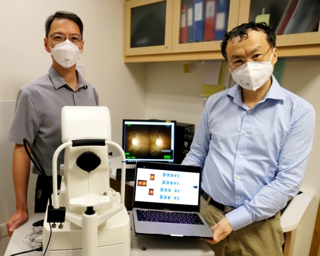
Scientists from Nanyang Technological University, Singapore (NTU Singapore), in collaboration with clinicians at Tan Tock Seng Hospital (TTSH), Singapore developed the AI-enabled method which uses algorithms to differentiate optic nerves with glaucoma from those that are normal. This is done by analysing ‘stereo fundus images’, multi-angle 2D images of the retina that are combined to form a 3D image.
When tested on stereo fundus images from TTSH patients undergoing expert examination, the AI method yielded an accuracy of 97 per cent in diagnosing glaucoma.
MORE FROM MEDICAL & HEALTHCARE
Glaucoma is usually asymptomatic until latter stages, when prognosis is poor. It is the principal cause of irreversible blindness worldwide and, in tandem with the rapid growth of the ageing population, is expected to affect 111.8 million people globally by 2040.
The automated glaucoma diagnosis method developed by NTU and TTSH, described in a study published in Methods in June 2021, could potentially be used in less developed areas where patients lack access to ophthalmologists, said the scientists.
Associate Professor Wang Lipo from the NTU School of Electrical and Electronic Engineering and lead author of the study said: “Through a combination of machine learning techniques, our team has developed a screening model that can diagnose glaucoma from fundus images, removing the need for ophthalmologists to take various clinical measurements…for diagnosis.
"The ease of use of our robust automated glaucoma diagnosis approach means that any healthcare practitioner could make use of the system to help in glaucoma screening. This will be especially helpful in geographical areas with less access to ophthalmologists.”
The automated glaucoma diagnostic system uses algorithms to analyse stereo fundus images taken as pairs by two cameras from different viewpoints. These 2D ‘left’ and ‘right’ images of the fundus help to form a 3D view when combined.
Using two images ensures that if one image is poor quality, the other image can usually compensate and the system can maintain its performance, the scientists said.
The set of algorithms is made up of a deep convolutional neural network and an attention-guided network. The former mimics the human brain’s biological process to adapt to learning new things, while the attention-guided network imitates the brain’s manner of selectively focusing on a few relevant features, such as the optic nerve head region in the fundus images.
The outputs from these two components are then fused together to generate the final prediction result.
To test their algorithms, the scientists reduced the resolution of 282 fundus images (70 glaucoma cases and 212 healthy cases) taken of TTSH patients during their eye screening, before training the algorithms with 70 per cent of the dataset.
To generate more training samples, the scientists also applied image augmentation – a technique that involves applying random but realistic transformations, such as image rotation – to increase the diversity of the dataset used to train the algorithms, which enhances the algorithms’ classification accuracy.
The team then tested their screening method on the remaining 30 per cent of the patient images and found that it had an accuracy of 97 per cent in correctly identifying glaucoma cases.
“Excellent and stable performance is especially important in medical diagnosis, and this study has shown that our model of combining deep convolutional neural network with attention mechanisms has resulted in a reliable and efficient AI-enabled screening approach for glaucoma,” said Prof Wang.


Labour pledge to tackle four key barriers in UK energy transition
I'm all for clarity and would welcome anyone who can enlighten me about what Labour's plans are for the size and scale of this Great British Energy....