Telescope technology helps microscope to image moving, living cells
Combining adaptive optics with a gentle use of light allows researchers to capture 3D videos of living cells inside organisms
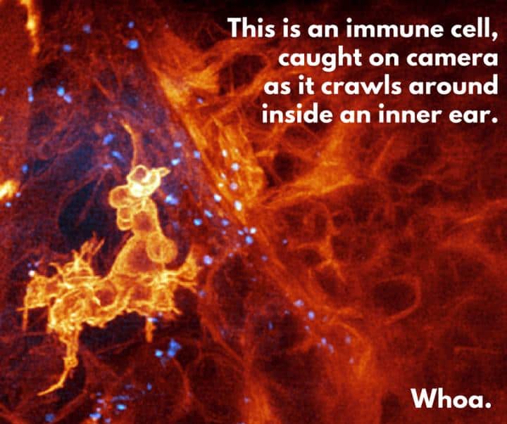
Watching cells is tricky. To determine how they behave in a living body, they must be viewed in an environment as close to natural as possible; and that means with the other cells that live alongside them. Light interacts differently with different shapes and substances, so conventional microscopy can only produce blurred and confusing images. Isolating the cells on a glass slide will not give information about their behaviour, and specialised microscopy using powerful forms of light can damage their delicate structure.
Researchers at the Howard Hughes Medical Institute in Virginia, working with Boston Children’s Hospital and Harvard Medical School, have devised a combination of technologies that produces startlingly sharp results.
The team, led by Dr Eric Betzig, co-winner of the 2014 Nobel Prize for chemistry for his role in developing super-resolved fluorescence microscopy, used a form of microscopy known as lattice light-sheet, which illuminates the subject by scanning a thin sheet of light repeatedly across living tissue at high speed to observe cells inside an embryonic zebrafish, suitable for this type of work because it has translucent skin.
Register now to continue reading
Thanks for visiting The Engineer. You’ve now reached your monthly limit of news stories. Register for free to unlock unlimited access to all of our news coverage, as well as premium content including opinion, in-depth features and special reports.
Benefits of registering
-
In-depth insights and coverage of key emerging trends
-
Unrestricted access to special reports throughout the year
-
Daily technology news delivered straight to your inbox


National Gas receives funding to develop Gravitricity underground hydrogen storage system
One single rock salt mine - Winsford - has 23 <i>MILLION </i>cubic metres of void and even allowing for 10% of that void set aside for hazardous waste...