Handheld probe images individual photoreceptors in infant eyes
Researchers have created a handheld probe that can image individual photoreceptors in the eyes of infants, a development that could help track early brain development in children.
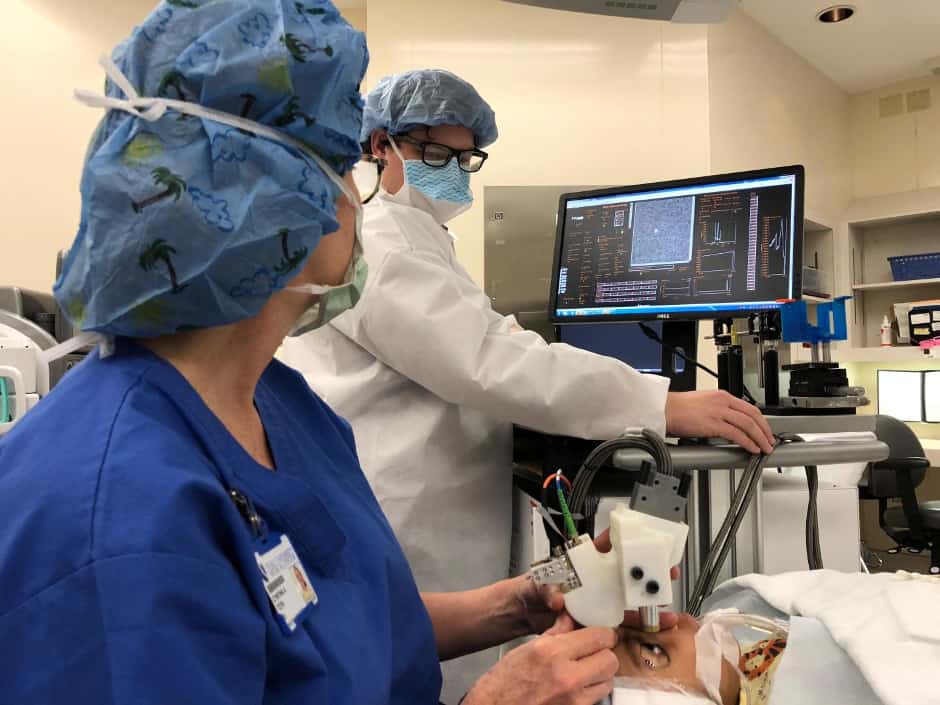
Based on adaptive optics, the technology from Duke University in North Carolina is expected to make it easier for doctors and scientists to observe these cells to diagnose eye diseases and make early detection of brain-related diseases and trauma.
Photoreceptors are specialised neurons comprising the light-sensing cells of the retina, an extension of the central nervous system located at the back of the eye. The retina sends signals to the brain via the optic nerve, which then processes the visual information. Studies have shown that neurodegenerative diseases, including Alzheimer's and Parkinson's, plus traumatic brain injuries, such as concussions, can alter the neuronal structures in the retina.
To study these neuronal structures, researchers commonly use an adaptive optics scanning laser ophthalmoscope (AOSLO), a non-invasive tool that provides higher image resolution than MRI.
Register now to continue reading
Thanks for visiting The Engineer. You’ve now reached your monthly limit of news stories. Register for free to unlock unlimited access to all of our news coverage, as well as premium content including opinion, in-depth features and special reports.
Benefits of registering
-
In-depth insights and coverage of key emerging trends
-
Unrestricted access to special reports throughout the year
-
Daily technology news delivered straight to your inbox


UK Enters ‘Golden Age of Nuclear’
The delay (nearly 8 years) in getting approval for the Rolls-Royce SMR is most worrying. Signifies a torpid and expensive system that is quite onerous...