
The Aston Brain Centre (ABC) will focus on child development disorders, including epilepsy, dyslexia, autism and attention deficit hyperactivity disorder (ADHD).
It will have a bespoke paediatric magnetoencephalography (MEG) scanner — thought to be the only one in Europe — developed jointly by clinicians at Aston University and Finnish manufacturer Elekta.
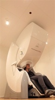
Unlike magnetic resonance imaging (MRI), which generates a large magnetic field of several Tesla around the body, MEG detects tiny, femto-Tesla magnetic fields naturally occurring in cells. Because of this sensitivity, it can distinguish similar cell types with different functions.
This makes it especially useful in surgical planning for difficult-to-treat brain conditions caused by a specific lesion, as ABC director Prof Paul Furlong explained.
‘If you had a lesion that was close to the motor cortex or speech and language areas, what the surgeon would normally do is remove the skull around those areas, wake you up and then stimulate those parts of the brain around the lesion to make sure that he doesn’t remove parts of the brain that are going to stop you from speaking or moving.
‘That’s something we’re doing non-invasively with MEG and it’s the ability to do this in younger and younger children that’s key here.’
Because of MEG’s sensitivity, the detector must be flush against the skull with little movement from the subject — a particular difficulty in children with behavioural problems. Their solution was to modify the architectures of the MEG cradle, as well as including bespoke software.
‘We track the head constantly during the recording and compensate for head movement using spherical harmonics, so if the child wriggles about it doesn’t matter — we can reconstitute the data so that we retain the spatial resolution.’
The ABC is also equipped with a new human brain tissue lab with specialist electrophysiology rigs. Lesion brain tissue resected from the aforementioned operations is kept alive on these rigs, bathed in cerebrospinal fluid, where pharmacologists can test various drugs on it and measure electrical response (as these patients have shown resistance to conventional treatment).
‘We go from measuring whole brain activity down to the individual neurons, or small clusters of neurons,’ Furlong said.


Nanogenerator consumes CO2 to generate electricity
Nice to see my my views being backed up by no less a figure than Sabine Hossenfelder https://youtu.be/QoJzs4fA4fo