Researchers at the University of California, Berkeley have developed a cell phone microscope that takes colour images of malaria parasites.
The prototype of the so-called CellScope could help take clinical microscopy out of specialised laboratories and into field settings for disease screening and diagnoses.
'The same regions of the world that lack access to adequate health facilities are, paradoxically, well served by mobile phone networks,' said Dan Fletcher, UC Berkeley associate professor of bioengineering and head of the research team developing the CellScope. 'We can take advantage of these mobile networks to bring low-cost, easy-to-use lab equipment out to more remote settings.'
The engineers attached compact microscope lenses to a holder fitted to a cell phone. Using samples of infected blood and sputum, the researchers were able to use the camera phone to capture bright field images of Plasmodium falciparum, the parasite that causes malaria in humans, and sickle-shaped red blood cells.
They were also able to take fluorescent images of Mycobacterium tuberculosis, the bacterial culprit that causes TB in humans. Moreover, the researchers showed that the TB bacteria could be automatically counted using image analysis software.
'The images can either be analysed on site or wirelessly transmitted to clinical centres for remote diagnosis,' said David Breslauer, a graduate student in the UC San Francisco/UC Berkeley Bioengineering Graduate Group. 'The system could be used to help provide early warning of outbreaks by shortening the time needed to screen, diagnose and treat infectious diseases.'
The researchers used filters to block out background light and to restrict the light source, a simple light-emitting diode (LED), to the 460 nanometre wavelength necessary to excite the green fluorescent dye in the TB-infected blood. Using an off-the-shelf phone with a 3.2 megapixel camera, they were able to achieve a spatial resolution of 1.2 micrometres. In comparison, a human red blood cell is about seven micrometres in diameter.
'A CellScope device with fluorescence could potentially also be used by patients undergoing chemotherapy who need to get regular blood counts,' said Fletcher. 'The patient could transmit from home the image or analysed data to a healthcare professional, reducing the number of clinical visits that are necessary.'
The CellScope developers have also been approached by experts in agriculture interested in using it to help diagnose diseases in crops. Instead of sending in a leaf sample to a lab for diagnosis, farmers could upload an image of the diseased leaf for analysis.
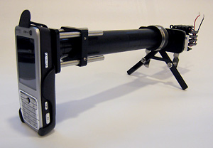
Schematic of the CellScope set up for fluorescent imaging. For bright field imaging, the two filters and LED are removed
The researchers are currently developing more robust prototypes of the CellScope in preparation for further field testing.


Nanogenerator consumes CO2 to generate electricity
Nice to see my my views being backed up by no less a figure than Sabine Hossenfelder https://youtu.be/QoJzs4fA4fo